Cell Meter™固定细胞和组织TUNEL细胞凋亡测定试剂盒*蓝色荧光* 货号22857-AAT Bioquest荧光染料

上海金畔生物科技有限公司代理AAT Bioquest荧光染料全线产品,欢迎访问AAT Bioquest荧光染料官网了解更多信息。

Cell Meter™固定细胞和组织TUNEL细胞凋亡测定试剂盒*蓝色荧光*

Cell Meter™固定细胞和组织TUNEL细胞凋亡测定试剂盒*蓝色荧光*

Cell Meter™固定细胞和组织TUNEL细胞凋亡测定试剂盒*蓝色荧光*    货号22857 货号 22857 存储条件 在零下15度以下保存, 避免光照
规格 25 Tests 价格 4992
Ex (nm) 411 Em (nm) 472
分子量 溶剂
产品详细介绍

简要概述

Cell Meter TUNEL细胞凋亡测定试剂盒是一种强大的工具,可方便地检测由DNA片段化引起的细胞凋亡。该测定是非放射性且快速的。TUNEL分析使用末端脱氧核苷酸转移酶(TdT)催化DEAC-dUTP在片段DNA的游离3′-羟基末端的掺入。通过荧光显微镜(AMC滤波片组)分析所得的DEAC标记的DNA。它的蓝色激发光可以方便地与GFP标记的靶标多路复用。荧光DEAC标记的核苷酸的直接掺入可明显减少检测步骤。该试剂盒经过优化,可检测固定细胞和福尔马林固定,石蜡包埋的组织切片中的凋亡。

点击查看光谱


适用仪器


流式细胞仪  
激发: 405nm激光
发射: 525/50nm
通道: Pacific Orange通道
荧光显微镜  
激发: 紫色滤波片组
发射: 紫色滤波片组
推荐孔板: 黑色透明


产品说明书

样品分析方案

概述

根据需要处理样品

在冰上用4%甲醛溶液固定细胞30分钟

在冰上用70%冰冷的乙醇透化细胞60分钟

将TdT染色溶液添加到样品中并在37°C下孵育60分钟

使用带有Violet滤光片组的荧光显微镜检测荧光强度 


工作溶液配制

TdT染色液

对于一项测试,混合以下试剂以使总体积为51 µL;
45 µL TdT反应缓冲液(组分D)
5 µL CoCl2(组分C)
0.5 µL TF5-dUTP(组分B)
0.5 µL TdT酶(组分A)。
注意:TdT染色溶液应立即使用。


操作步骤

细胞染色方案

以下方案可用作参考,实际情况应根据需要进行调整。

  1. 根据需要处理样品。
  2. 用您选择的缓冲液(例如含Ca +2和Mg +2的 PBS)洗涤样品。
  3. 通过在PBS中加入100 µL 4%多聚甲醛固定样品,并在冰上孵育30分钟。
  4. 除去固定液并用PBS洗涤样品。
  5. 向样品中加入100 µL 70%的冰冷乙醇,并在冰上孵育60分钟。
    注意:样品在使用前可以在-20°C下保存几天。
  6. 除去酒精并用PBS洗涤细胞。
    注意:对于阳性对照,将固定样品与2-5 µg / mL DNAse在含有Ca +2和Mg +2的PBS中于37°C 孵育60分钟。除去DNAse并彻底清洗细胞,并继续进行其余操作。
  7. 向样品中加入50µL TdT染色溶液,并在37°C下孵育60至120分钟。
  8. 除去TdT工作溶液,并用PBS洗涤样品。
  9. 将样品重悬于PBS中,并使用流式细胞仪使用525/50 nm滤光片(Pacific Orange通道)或带有紫色滤光片组的荧光显微镜检测荧光强度。 


组织染色方案

以下方案可用作参考,实际应根据需要进行调整。
去石蜡和水化

  1. 通过将载玻片在Coplin广口瓶中在室温下浸入新鲜的二甲苯中5分钟,从而对组织切片(附着在载玻片上)进行脱蜡。再重复一次。(共2次洗涤)
  2. 将载玻片在室温下在Coplin广口瓶中浸入100%乙醇中5分钟,以洗涤样品。
  3. 通过在室温下将载玻片浸入各种浓度的酒精(100、95、85、70、50%)中,分别在室温下浸泡5分钟,从而对样品进行水化处理。
  4. 在室温下将载玻片浸入0.85%NaCl中5分钟,以洗涤样品。
  5. 在室温下将载玻片浸入PBS中5分钟,以洗涤样品。重复一遍。(共2次洗涤) 


固定

  1. 通过在室温下将载玻片浸入PBS中的4%多聚甲醛溶液中15-20分钟来固定组织切片。
  2. 在室温下将载玻片浸入PBS中5分钟,以洗涤样品。重复一遍。(共2次洗涤)
  3. 除去液体,然后将载玻片放在平坦的地方。用100 µL 20 µg / mL蛋白酶K溶液处理组织切片。(添加至足够覆盖整个组织表面。在室温下孵育载玻片10分钟。)
  4. 在室温下将载玻片浸入PBS中5分钟,以洗涤样品。
  5. 通过在室温下将载玻片浸入PBS中的4%多聚甲醛溶液中15-20分钟来固定组织切片。
  6. 在室温下将载玻片浸入PBS中5分钟,以洗涤样品。重复一遍。(共2次洗涤) 


染色

  1. 可选:对于阳性对照,将固定的样品与2-5 µg / mL的DNAse在含有Ca +2和Mg +2的PBS中于37°C 孵育60分钟。除去DNAse,并用PBS彻底清洗细胞,并继续进行其余操作。
  2. 向样品中加入50 µL TdT染色溶液,并在37°C下孵育60至120分钟。
  3. 除去TdT工作溶液,并用PBS洗涤样品。
  4. 加入封固剂,并用带紫色滤光片组的荧光显微镜检测荧光强度。 


图示

Cell Meter™固定细胞和组织TUNEL细胞凋亡测定试剂盒*蓝色荧光*    货号22857

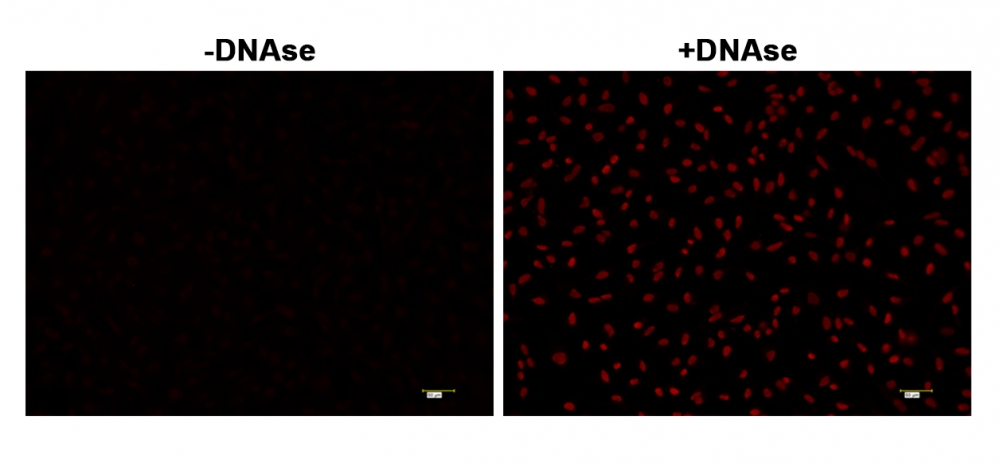
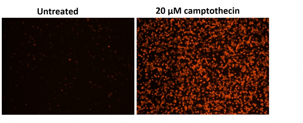
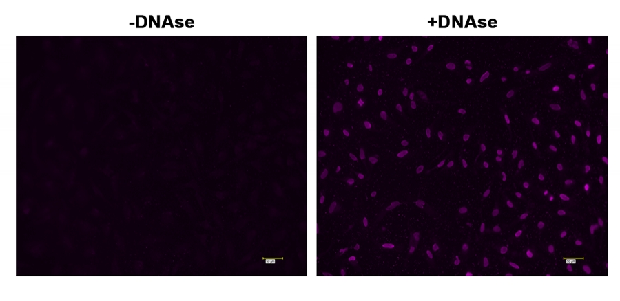
图1.用   HeLa细胞进行TUNEL分析的荧光图像。 固定HeLa细胞,并在37°C下分别使用和不使用DNAse处理60分钟。然后将细胞用Cell Meter™固定细胞和组织TUNEL细胞凋亡测定试剂盒(Cat#22855)染色。DNA链断裂在DNAse处理的细胞中表现出强烈的荧光染色。使用紫色滤光片组在荧光显微镜下检测信号。  


参考文献

A Novel Theranostic Combination of Near-infrared Fluorescence Imaging and Laser Irradiation Targeting c-KIT for Gastrointestinal Stromal Tumors.
Authors: Fujimoto, Shota and Muguruma, Naoki and Okamoto, Koichi and Kurihara, Takeshi and Sato, Yasushi and Miyamoto, Yoshihiko and Kitamura, Shinji and Miyamoto, Hiroshi and Taguchi, Takahiro and Tsuneyama, Koichi and Takayama, Tetsuji
Journal: Theranostics (2018): 2313-2328

Multicomponent Synthesis of Some Molecular Hybrid Containing Thiazole Pyrazole as Apoptosis Inducer.
Authors: Kumar, Parvin and Duhan, Meenakshi and Kadyan, Kulbir and Bhardwaj, Jitender Kumar and Saraf, Priyanka and Mittal, Meenu
Journal: Drug research (2018): 72-79

[Salvianolate protects H9c2 cells from hypoxia/reoxygenation injury-induced apoptosis by attenuating mitochondrial DNA oxidative damage].
Authors: Yue, R C and Yang, X L and Zhang, R Y and Liu, S and Liu, J and Zeng, J and Liang, H and Wang, W and Hu, H X and Zeng, C Y
Journal: Zhonghua xin xue guan bing za zhi (2017): 57-63

Caspase 3 as a therapeutic target for regulation of intervertebral disc degeneration in rabbits.
Authors: Sudo, Hideki and Minami, Akio
Journal: Arthritis and rheumatism (2011): 1648-57

Induction of apoptosis and inhibition of PI3K/Akt pathway in PC-3 and LNCaP prostate cancer cells by ethanolic neem leaf extract.
Authors: Gunadharini, Dharmalingam N and Elumalai, Perumal and Arunkumar, Ramachandran and Senthilkumar, Kalimuthu and Arunakaran, Jagadeesan
Journal: Journal of ethnopharmacology (2011): 644-50

[Effect of 5-aminolevulinic acid-mediated photodynamic therapy on human gastric cancer xenografts in nude mice in vivo].
Authors: Zhou, Guang-jun and Huang, Zong-hai and Yu, Jin-long and Li, Zhou and Ding, Lian-shu
Journal: Zhonghua wei chang wai ke za zhi = Chinese journal of gastrointestinal surgery (2008): 580-3

[Effect of phenylhexyl isothiocyanate on inducing apoptosis of multiple myeloma cells in vitro].
Authors: Lu, Quan-Yi and Wang, Zhao and Liu, De-Long
Journal: Zhongguo shi yan xue ye xue za zhi (2008): 89-92

[Effect of qihong capsule in inhibiting cell apoptosis induced by Coxsackie virus B].
Authors: Song, Xiao-dong and Wang, Lun and Ji, Bo
Journal: Zhongguo Zhong xi yi jie he za zhi Zhongguo Zhongxiyi jiehe zazhi = Chinese journal of integrated traditional and Western medicine (2005): 511-5

Dual excitation multi- fluorescence flow cytometry for detailed analyses of viability and apoptotic cell transition.
Authors: Mazzini, G and Ferrari, C and Erba, E
Journal: European journal of histochemistry : EJH (2003): 289-98

Co-localization of active caspase-3 and DNA fragmentation (TUNEL) in normal and hyperthermia-induced abnormal mouse development.
Authors: Mirkes, P E and Little, S A and Umpierre, C C
Journal: Teratology (2001): 134-43

说明书
Cell Meter™固定细胞和组织TUNEL细胞凋亡测定试剂盒*蓝色荧光*.pdf

Cell Meter Caspase 8活性细胞凋亡检测试剂盒 蓝色荧光 货号22812-AAT Bioquest荧光染料

上海金畔生物科技有限公司代理AAT Bioquest荧光染料全线产品,欢迎访问AAT Bioquest荧光染料官网了解更多信息。

Cell Meter Caspase 8活性细胞凋亡检测试剂盒 蓝色荧光

Cell Meter Caspase 8活性细胞凋亡检测试剂盒 蓝色荧光

Cell Meter Caspase 8活性细胞凋亡检测试剂盒 蓝色荧光     货号22812 货号 22812 存储条件 在零下15度以下保存, 避免光照
规格 200 tests 价格 2604
Ex (nm) 341 Em (nm) 441
分子量 溶剂
产品详细介绍

简要概述

Cell Meter Caspase 8活性细胞凋亡检测试剂盒是美国AAT Bioquest生产的用于细胞凋亡检测的试剂盒,Cell Meter检测试剂盒是一类用于检测细胞功能的系列工具,包括细胞活性、细胞毒性、细胞凋亡、细胞膜电位以及细胞周期等方面的指标。每种检测方案均能提供不同荧光颜色的检测方案。这些高效的检测方案为从多角度研究细胞功能活动提供了一种十分有效的方法。Cell Meter Caspase 8活性细胞凋亡检测试剂盒 *蓝色荧光* 是通过测量Caspase 8的活性而用于细胞凋亡检测的。Caspase 8 是一种Caspase蛋白,CASP8基因所编码。在神经元变性疾病中,例如亨廷顿综合症,caspase 8扮演着重要角色。Caspase 8已被证明具有底物选择性,其肽基序为Ile-Glu-Thr-Asp (IETD)。该试剂盒使用(Ac-IETD)2-AMC作为荧光指示器来检测caspas-8的活性。通过caspas-8的切割AMC肽产生强蓝色荧光(eX/eM = 350/450 NM)。这种特性使得试剂盒能适合于DAPI滤光器。本试剂盒提供所有必需组分和最佳操作流程。该实验结果稳定,快速且适应高通量筛选。它不仅能够定量凋亡细胞中caspas-8的活性还能筛选caspas-8的抑制剂。金畔生物是AAT Bioquest的中国代理商,为您提供最优质的Cell Meter Caspase 8活性细胞凋亡检测试剂盒。 

点击查看光谱


适用仪器


荧光酶标仪  
激发: 370nm
发射: 450nm
cutoff: 420nm
推荐孔板: 黑色透明
读取模式: 顶或底读模式

产品说明书

样品实验方案

简要概述

1.用测试化合物(100μL/孔/ 96孔板或25μL/孔/ 384孔板)制备细胞
2.加入等体积的Caspase 8工作溶液(100μL/孔/ 96孔板或25μL/孔/ 384孔板)
3.在室温下孵育30-60分钟
4.监测Ex / Em = 370/450 nm处的荧光增加(截止= 420 nm)


工作溶液配制

将50μLCaspase8底物(组分A)加入10mL测定缓冲液(组分B)中并充分混合以制备Caspase 8工作溶液。 避光。 注意:Caspase 8工作液不稳定,请及时使用。有关细胞样品制备的指南,请点击查看


操作步骤

1.通过将10μL/孔的10X测试化合物(96孔板)或5μL/孔的5X测试化合物(384孔板)加入PBS或所需缓冲液中来处理细胞。对于空白孔(没有细胞的培养基),加入相同量的化合物缓冲液。

2.将细胞板在5%CO 2,37℃培养箱中孵育所需的一段时间(对于用星形孢菌素处理的Jurkat细胞4-6小时)以诱导细胞凋亡。

3.加入100μL/孔(96孔板)或25μL/孔(384孔板)的Caspase 8工作溶液。

4.将Caspase 8工作溶液板在室温下孵育30至60分钟,避光。注意:如果需要,在室温下加入Caspase 8工作溶液10分钟前,向选定的样品中加入1μL的1 mM Ac-IETD-CHO caspase 8抑制剂,以确认对caspase 8样活性的抑制作用。

5.以800rpm离心细胞板(特别是对于非贴壁细胞)2分钟(制动关闭)。

6.用荧光酶标仪在Ex / Em = 370 / 450nm(截止= 420nm)下监测荧光增加。


数据分析

Cell Meter Caspase 8活性细胞凋亡检测试剂盒 蓝色荧光     货号22812

图1.使用Cell Meter Caspase 8活性细胞凋亡检测试剂盒检测Jurkat细胞中的caspase 8活性*蓝色荧光*。 在Costar黑壁/透明底96孔板中以相同的一天以200,000个细胞/90μL/孔接种Jurkat细胞。 用或不用1μM星形孢菌素处理细胞5小时。 加入半胱天冬酶8工作溶液(100μL/孔)并在室温下温育30分钟。 使用FlexStation 酶标仪(Molecular Devices)在Ex / Em = 370 / 450nm(截止值= 420nm)下测量荧光强度。


参考文献

Angiopoietins Modulate Survival, Migration, and the Components of the Ang-Tie2 Pathway of Chronic Lymphocytic Leukaemia (CLL) Cells In Vitro
Authors: Luis Mario Aguirre Palma, Hanna Flamme, Iris Gerke, Karl-Anton Kreuzer
Journal: Cancer Microenvironment (2016): 13–26

Hypoxia regulates TRAIL sensitivity of colorectal cancer cells through mitochondrial autophagy.
Authors: Gertrud Knoll, Sebastian Bittner, Maria Kurz, Jonathan Jantsch, Martin Ehrenschwender
Journal: Oncotarget (2016)

microRNA-186 inhibits cell proliferation and induces apoptosis in human esophageal squamous cell carcinoma by targeting SKP2
Authors: Wei He, Jianfang Feng, Yan Zhang, Yuanyuan Wang, Wenqiao Zang, Guoqiang Zhao
Journal: Laboratory Investigation (2016): 317–324

Promotion of osteointegration under diabetic conditions by tantalum coating-based surface modification on 3-dimensional printed porous titanium implants
Authors: Lin Wang, Xiaofan Hu, Xiangyu Ma, Zhensheng Ma, Yang Zhang, Yizhao Lu, Xiang Li, Wei Lei, Yafei Feng
Journal: Colloids and Surfaces B: Biointerfaces (2016): 440–452

Role of delta-like ligand-4 in chemoresistance against docetaxel in MCF-7 cells
Authors: Q Wang, Y Shi, HJ Butler, J Xue, G Wang, P Duan, H Zheng
Journal: Human & Experimental Toxicology (2016): 0960327116650006

Glucose promotes cell proliferation, glucose uptake and invasion in endometrial cancer cells via AMPK/mTOR/S6 and MAPK signaling
Authors: Jianjun Han, Lu Zhang, Hui Guo, Weiya Z Wysham, Dario R Roque, Adam K Willson, Xiugui Sheng, Chunxiao Zhou, Victoria L Bae-Jump
Journal: Gynecologic oncology (2015): 668–675

IL-7 abrogates the immunosuppressive function of human double-negative T cells by activating Akt/mTOR signaling
Authors: Andrea Allgäuer, Elisabeth Schreiner, Fulvia Ferrazzi, Arif B Ekici, Armin Gerbitz, Andreas Mackensen, Simon Völkl
Journal: The Journal of Immunology (2015): 3139–3148

Insulin improves osteogenesis of titanium implants under diabetic conditions by inhibiting reactive oxygen species overproduction via the PI3K-Akt pathway
Authors: Lin Wang, Xiong Zhao, Bo-yuan Wei, Yi Liu, Xiang-yu Ma, Jian Wang, Peng-chong Cao, Yang Zhang, Ya-bo Yan, Wei Lei
Journal: Biochimie (2015): 85–93

LGL1 modulates proliferation, apoptosis, and migration of human fetal lung fibroblasts
Authors: Hui Zhang, Neil B Sweezey, Feige Kaplan
Journal: American Journal of Physiology-Lung Cellular and Molecular Physiology (2015): L391–L402

t-BHQ Provides Protection against Lead Neurotoxicity via Nrf2/HO-1 Pathway
Authors: Fang Ye, Xiaoyi Li, Lili Li, Jing Yuan, Jun Chen
Journal: Oxidative medicine and cellular longevity (2015)


相关产品

产品名称 货号
Cell Meter Caspase 8活性细胞凋亡检测试剂盒 绿色荧光 Cat#22798
Cell Meter Caspase 8活性细胞凋亡检测试剂盒 红色荧光 Cat#22816
Cell Meter Caspase 9活性细胞凋亡检测试剂盒 红色荧光 Cat#22817

说明书
Cell Meter Caspase 8活性细胞凋亡检测试剂盒 蓝色荧光 .pdf

Cell Meter 磷脂酰丝氨酸凋亡检测试剂盒 橙色荧光,适合酶标仪检测 货号22794-AAT Bioquest荧光染料

上海金畔生物科技有限公司代理AAT Bioquest荧光染料全线产品,欢迎访问AAT Bioquest荧光染料官网了解更多信息。

Cell Meter 磷脂酰丝氨酸凋亡检测试剂盒 橙色荧光,适合酶标仪检测

Cell Meter 磷脂酰丝氨酸凋亡检测试剂盒 橙色荧光,适合酶标仪检测

Cell Meter 磷脂酰丝氨酸凋亡检测试剂盒 橙色荧光,适合酶标仪检测     货号22794 货号 22794 存储条件 在2-8度冷藏保存, 避免光照
规格 100 Tests 价格 2604
Ex (nm) 554 Em (nm) 578
分子量 溶剂
产品详细介绍

简要概述

Cell Meter检测试剂盒是一类用于检测细胞功能的系列工具,该特定试剂盒旨在通过测量磷脂酰丝氨酸(PS)的转运来检测细胞凋亡。在细胞凋亡中,PS转移到质膜的外部小叶。磷脂酰丝氨酸在细胞表面的出现是细胞凋亡的初始/中间阶段的通用指标,可以在观察形态变化之前进行检测。该试剂盒使用我们专有的橙色荧光Apopxin PS传感器,该传感器特异性结合PS,其亲和力远高于Annexin V(Kd <10 nM)。该试剂盒中使用的PS传感器与膜PS结合后具有橙色荧光。它的光谱性质几乎与Cy3®或AlexaFluor®555的光谱性质相同,使其易于用配备有Cy3®或AlexaFluor®555光源和滤光片的普通荧光仪器(Cy3®或AlexaFluor®555分别是GE Healthcare和Invitrogen的商标)进行实验。由于它对PS的亲和力大大增强,因此该试剂盒比仅与显微镜或流式细胞仪平台一起使用的其他基于膜联蛋白V的商业化凋亡试剂盒更为强大。除显微镜和流式细胞仪平台外,该试剂盒还可与荧光酶标仪一起使用。

点击查看细胞样品配制


适用仪器


荧光酶标仪  
激发: 540nm
发射: 590nm
cutoff: 570nm
推荐孔板: 黑色透明
读取模式: 底读模式

产品说明书

样品实验方案

简要概述

  1. 用测试化合物制备细胞(100 µL /孔/ 96孔板或25 µL /孔/ 384孔板)
  2. 加入等量的Apopxin Orange工作溶液
  3. 在室温下孵育1小时
  4. 在Ex / Em = 540/590 nm(截止= 570 nm)或使用Cy3滤光片的荧光显微镜下检测荧光强度(底部读取模式)


溶液配制

工作溶液配制

将10μL的100X Apopxin 橙色(组分A)添加到1 mL的测定缓冲液(组分B)中,并充分混合以制成Apopxin Orange工作溶液。 注意: 100μLApopxin Orange工作溶液足以用于一个孔。现用现配避免长时间放置。


操作步骤

1.通过向PBS或所需的缓冲液中加入10ul /孔(96孔板)或2.5 µL /孔(384孔板)的10X测试化合物储备溶液来处理细胞。 对于空白孔(不含细胞的培养基),添加相同量的化合物缓冲液。

2.将细胞板在5%CO2、37°C的培养箱中孵育(对于喜树碱处理过的Jurkat细胞,需要4-6小时)以诱导凋亡。

3.在每个孔中添加100 µL /孔(96孔板)或25 µL /孔(384孔板)的Apopxin Orange工作溶液。

4.在避光条件下,于室温下孵育细胞板至少1小时。

5.以800 rpm的速度离心细胞板(特别是非贴壁细胞)2分钟(制动)。

6.使用荧光酶标仪(Ext / Em = 540/590 nm(截止= 570 nm))或使用带有Cy3®滤光片的荧光显微镜对图像进行荧光强度检测。


图示

Cell Meter 磷脂酰丝氨酸凋亡检测试剂盒 橙色荧光,适合酶标仪检测     货号22794

图1.用Cell Meter 磷脂酰丝氨酸细胞凋亡检测试剂盒染色的Costar黑色96孔板中Jurkat细胞的荧光图像。 不使用(左)/使用20 µM喜树碱(右)处理Jurkat细胞5小时。 使用具有通道的荧光显微镜测量荧光强度。


参考文献

Physiological effects of the herbicide glyphosate on the cyanobacterium Microcystis aeruginosa
Authors: Wu, Liang and Qiu, Zhihao and Zhou, Ya and Du, Yuping and Liu, Chaonan and Ye, Jing and Hu, Xiaojun
Journal: Aquatic Toxicology (2016): 72–79

Tumor-selective mitochondrial network collapse induced by atmospheric gas plasma-activated medium.
Authors: Saito, Kosuke and Asai, Tomohiko and Fujiwara, Kyoko and Sahara, Junki and Koguchi, Haruhisa and Fukuda, Noboru and Suzuki-Karasaki, Miki and Soma, Masayoshi and Suzuki-Karasaki, Yoshihiro
Journal: Oncotarget (2016)

Inhibition of malignant phenotypes of human osteosarcoma cells by a gene silencer, a pyrrole–imidazole polyamide, which targets an E-box motif
Authors: Taniguchi, Masashi and Fujiwara, Kyoko and Nakai, Yuji and Ozaki, Toshinori and Koshikawa, Nobuko and Toshio, Kojima and Kataba, Motoaki and Oguni, Asako and Matsuda, Hiroyuki and Yoshida, Yukihiro and others
Journal: FEBS open bio (2014): 328–334

Detection of apoptosis based on the interaction between annexin V and phosphatidylserine
Authors: Liu T, Zhu W, Yang X, Chen L, Yang R, Hua Z, Li G.
Journal: Anal Chem (2009): 2410

Evaluation of cell surface expression of phosphatidylserine in ovarian carcinoma effusions using the annexin-V/7-AAD assay: clinical relevance and comparison with other apoptosis parameters
Authors: Dong HP, Holth A, Kleinberg L, Ruud MG, Elstr and MB, Trope CG, Davidson B, Risberg B.
Journal: Am J Clin Pathol (2009): 756

Mobilization of lysosomal calcium regulates the externalization of phosphatidylserine during apoptosis
Authors: Mirnikjoo B, Balasubramanian K, Schroit AJ.
Journal: J Biol Chem (2009): 6918

Peptidic targeting of phosphatidylserine for the MRI detection of apoptosis in atherosclerotic plaques
Authors: Burtea C, Laurent S, Lancelot E, Ballet S, Murariu O, Rousseaux O, Port M, V and er Elst L, Corot C, Muller RN.
Journal: Mol Pharm (2009): 1903

Suicidal membrane repair regulates phosphatidylserine externalization during apoptosis
Authors: Mirnikjoo B, Balasubramanian K, Schroit AJ.
Journal: J Biol Chem (2009): 22512

Trivalent methylated arsenical-induced phosphatidylserine exposure and apoptosis in platelets may lead to increased thrombus formation
Authors: Bae ON, Lim KM, Noh JY, Chung SM, Kim SH, Chung JH.
Journal: Toxicol Appl Pharmacol (2009): 144

Discovery of a phosphatidylserine-recognizing peptide and its utility in molecular imaging of tumour apoptosis
Authors: Thapa N, Kim S, So IS, Lee BH, Kwon IC, Choi K, Kim IS.
Journal: J Cell Mol Med (2008): 1649

说明书
Cell Meter 磷脂酰丝氨酸凋亡检测试剂盒 橙色荧光,适合酶标仪检测 .pdf

胱天蛋白酶Caspase抑制剂Z-VAD-FMK 货号13300-AAT Bioquest荧光染料

上海金畔生物科技有限公司代理AAT Bioquest荧光染料全线产品,欢迎访问AAT Bioquest荧光染料官网了解更多信息。

胱天蛋白酶Caspase抑制剂Z-VAD-FMK

胱天蛋白酶Caspase抑制剂Z-VAD-FMK

胱天蛋白酶Caspase抑制剂Z-VAD-FMK    货号13300 货号 13300 存储条件 在零下15度以下保存, 避免光照
规格 1 mg 价格 1200
Ex (nm) Em (nm)
分子量 467.49 溶剂 DMSO
产品详细介绍

简要概述

Z-VAD-FMK是细胞可渗透的不可逆半胱天冬酶抑制剂,与半胱天冬酶蛋白的催化位点结合,可抑制凋亡的诱导。为了抑制细胞凋亡,应在诱导细胞凋亡的同时添加Z-VAD-FMK。该肽在天冬氨酸的P1位置被O-甲基化,提供增强的稳定性和更高的细胞通透性。FMK衍生的肽可作为有效的不可逆抑制剂,而不会增加细胞毒性作用。金畔生物是AAT Bioquest的中国代理商,为您提供最优质的Z-VAD-FMK。 

产品说明书

样品实验方案

以下是我们推荐的方案,仅提供指导。具体实验应根据您的特定需求进行修改。


溶液配制

储备溶液配制

Z-VAD-FMK储备溶液配制:在Z-VAD-FMK中添加适量的DMSO,制成2-5 mM Z-VAD-FMK储备液。注意:将未使用的Z-VAD-FMK储备溶液分装在-20°C下。


操作步骤

  1. 根据需要处理样品以诱导凋亡。
  2. 添加Z-VAD-FMK。
    注意:我们建议使用最终浓度为10-100 µM的Z-VAD-FMK抑制胱天蛋白酶,例如对caspase-8抑制50 µM。
    注意:孵育的最佳时间和浓度需要通过实验确定。
  3. 根据需要对样品染色。
  4. 用适当的仪器检测荧光强度。 


图示


胱天蛋白酶Caspase抑制剂Z-VAD-FMK    货号13300

图1.用半胱天冬酶抑制剂Z-VAD-FMK(Cat#13300)抑制Jurkat细胞中的胱天蛋白酶活性。当天,将Jurkat细胞以200,000个细胞/ 90 µL /孔的密度接种在Costar黑色96孔板中。用1 µM星形孢菌素处理细胞5小时。加入Z-VAD-FMK(50 uM)并与星形孢菌素共孵育。用Cell Meter Caspase 8活性细胞凋亡检测试剂盒(Cat#22816)检测caspase-8活性。用Flexstation 3荧光酶标仪(Molecular Devices)在Ex / Em = 540 / 620nm(截止= 610nm)下测量荧光强度。


参考文献

Identification of active caspases using affinity-based probes.
Authors: McStay, Gavin P and Green, Douglas R
Journal: Cold Spring Harbor protocols (2014): 856-60

Induction of apoptosis in human renal cell carcinoma cells by vitamin E succinate in caspase-independent manner.
Authors: Wu, Xiu-Xian and Kakehi, Yoshiyuki and Jin, Xing-Hua and Inui, Masashi and Sugimoto, Mikio
Journal: Urology (2009): 193-9

Induction of apoptosis in renal tubular cells by histone deacetylase inhibitors, a family of anticancer agents.
Authors: Dong, Guie and Wang, Lysa and Wang, Cong-Yi and Yang, Tianxin and Kumar, M Vijay and Dong, Zheng
Journal: The Journal of pharmacology and experimental therapeutics (2008): 978-84

Metformin induces apoptosis of pancreatic cancer cells.
Authors: Wang, Luo-Wei and Li, Zhao-Shen and Zou, Duo-Wu and Jin, Zhen-Dong and Gao, Jun and Xu, Guo-Ming
Journal: World journal of gastroenterology (2008): 7192-8

Apoptosis of gastric cancer cell line MKN45 by photodynamic treatment with photofrin.
Authors: Takahira, Kenichiro and Sano, Munetaka and Arai, Hajime and Hanai, Hiroyuki
Journal: Lasers in medical science (2004): 89-94

Non-caspase-mediated apoptosis contributes to the potent cytotoxicity of the enediyne antibiotic lidamycin toward human tumor cells.
Authors: Wang, Zhen and He, Qiyang and Liang, Yunyan and Wang, Daishu and Li, Yi Yang and Li, Diandong
Journal: Biochemical pharmacology (2003): 1767-75

Androgen induced cell death in SHSY5Y neuroblastoma cells expressing wild-type and spinal bulbar muscular atrophy mutant androgen receptors.
Authors: Grierson, A J and Shaw, C E and Miller, C C
Journal: Biochimica et biophysica acta (2001): 13-20

Critical role for caspases 3 and 8 in neutrophil but not eosinophil apoptosis.
Authors: Daigle, I and Simon, H U
Journal: International archives of allergy and immunology (2001): 147-56

说明书
胱天蛋白酶Caspase抑制剂Z-VAD-FMK.pdf

Cell Meter Caspase 9活性细胞凋亡检测试剂盒 蓝色荧光 货号22813-AAT Bioquest荧光染料

上海金畔生物科技有限公司代理AAT Bioquest荧光染料全线产品,欢迎访问AAT Bioquest荧光染料官网了解更多信息。

Cell Meter Caspase 9活性细胞凋亡检测试剂盒 蓝色荧光

Cell Meter Caspase 9活性细胞凋亡检测试剂盒 蓝色荧光

Cell Meter Caspase 9活性细胞凋亡检测试剂盒 蓝色荧光     货号22813 货号 22813 存储条件 在零下15度以下保存, 避免光照
规格 200 Tests 价格 2604
Ex (nm) 341 Em (nm) 441
分子量 溶剂
产品详细介绍

简要概述

Cell Meter Caspase 9活性细胞凋亡检测试剂盒是美国AAT Bioquest生产的用于细胞凋亡检测的试剂盒,Cell Meter检测试剂盒是一类用于检测细胞功能的系列工具,包括细胞活性、细胞毒性、细胞凋亡、细胞膜电位以及细胞周期等方面的指标。每种检测方案均能提供不同荧光颜色的检测方案。这些高效的检测方案为从多角度研究细胞功能活动提供了一种十分有效的方法。Cell Meter Caspase 9活性细胞凋亡检测试剂盒 *蓝色荧光* 是通过测量Caspase 9的活性而用于细胞凋亡检测的。Caspase 9是CED-3亚家族中的一员。活化的Caspase 9能切割下游caspases,例如caspase-3,-6和-7,起始Caspase级联反应。在正常发育的中枢神经系统中,活化的Caspase 9是凋亡所必需的。Caspase 9具有底物的选择性,其识别底物基序为Leu-Glu-His-Asp (LEHD)。本试剂盒利用Ac-LEHD-AMC作为荧光指示器来检测caspas-9的活性。通过caspas-9的切割AMC产生强蓝色荧光,eX/eM = 350/450 nm。这种特性使得试剂盒能适合于DAPI滤光器。本试剂盒提供所有必需组分。该实验稳定、快速且适应高通量筛选的需求。它不仅能够定量凋亡细胞中caspas-9的活性还能筛选caspas-9的抑制剂。很多实验室利用本试剂盒进行高通量抑制剂的筛选。金畔生物是AAT Bioquest的中国代理商,为您提供最优质的Cell Meter Caspase 9活性细胞凋亡检测试剂盒。 

点击查看光谱


适用仪器


荧光酶标仪  
激发: 375nm
发射: 435nm
cutoff: 420nm
推荐孔板: 黑色透明
读取模式: 顶或底读模式

产品说明书

样品实验分析

简要概述

用测试化合物(100μL/孔/ 96孔板或25μL/孔/ 384孔板)制备细胞
加入等体积的Caspase 9工作溶液(100μL/孔/ 96孔板或25μL/孔/ 384孔板)
在室温下孵育30-60分钟
监测Ex / Em = 375/435 nm处的荧光(截止= 420 nm)


工作溶液配制

将50μLCaspase9底物(组分A)加入10mL测定缓冲液(组分B)中并充分混合以制备Caspase 9工作溶液。 避光。有关细胞样品制备的指南,请点击查看

操作步骤

1.通过将10μL/孔的10X测试化合物(96孔板)或5μL/孔的5X测试化合物(384孔板)加入PBS或所需缓冲液中来处理细胞。对于空白孔(没有细胞的培养基),加入相同量的化合物缓冲液。

2.将细胞板在5%CO2,37℃培养箱中孵育所需的一段时间(对于用星形孢菌素处理的Jurkat细胞,4-6小时)以诱导细胞凋亡。

3.加入100μL/孔(96孔板)或25μL/孔(384孔板)的Caspase 9工作溶液。

4.将Caspase 9工作溶液板在室温下孵育至少1小时,避光。注意:如果需要,在室温下加入Caspase 9工作溶液前10分钟,向选定的样品中加入1μL的1 mM Ac-LEHD-CHO caspase 9抑制剂,以确认对胱天蛋白酶9样活性的抑制作用。

5.将细胞板(特别是非贴壁细胞)以800rpm离心2分钟。

6.用荧光酶标仪在Ex / Em = 375 / 435nm(截止= 420nm)下监测荧光强度。


数据分析

Cell Meter Caspase 9活性细胞凋亡检测试剂盒 蓝色荧光     货号22813

图1.使用Cell Meter Caspase 9活性细胞凋亡检测试剂盒检测Jurkat细胞中的Caspase 9活性 蓝色荧光 。 将Jurkat细胞以300,000个细胞/90μL/孔接种在Costar黑色壁/透明底96孔板中。 用或不用1μM星形孢菌素处理细胞4小时。 加入半胱天冬酶9工作溶液(100μL/孔)并在室温下温育1小时。 使用FlexStation 酶标仪(Molecular Devices)在Ex / Em = 375 / 435nm(截止值= 420nm)下测量荧光强度。


参考文献

Angiopoietins Modulate Survival, Migration, and the Components of the Ang-Tie2 Pathway of Chronic Lymphocytic Leukaemia (CLL) Cells In Vitro
Authors: Luis Mario Aguirre Palma, Hanna Flamme, Iris Gerke, Karl-Anton Kreuzer
Journal: Cancer Microenvironment (2016): 13–26

Hypoxia regulates TRAIL sensitivity of colorectal cancer cells through mitochondrial autophagy.
Authors: Gertrud Knoll, Sebastian Bittner, Maria Kurz, Jonathan Jantsch, Martin Ehrenschwender
Journal: Oncotarget (2016)

microRNA-186 inhibits cell proliferation and induces apoptosis in human esophageal squamous cell carcinoma by targeting SKP2
Authors: Wei He, Jianfang Feng, Yan Zhang, Yuanyuan Wang, Wenqiao Zang, Guoqiang Zhao
Journal: Laboratory Investigation (2016): 317–324

Promotion of osteointegration under diabetic conditions by tantalum coating-based surface modification on 3-dimensional printed porous titanium implants
Authors: Lin Wang, Xiaofan Hu, Xiangyu Ma, Zhensheng Ma, Yang Zhang, Yizhao Lu, Xiang Li, Wei Lei, Yafei Feng
Journal: Colloids and Surfaces B: Biointerfaces (2016): 440–452

Role of delta-like ligand-4 in chemoresistance against docetaxel in MCF-7 cells
Authors: Q Wang, Y Shi, HJ Butler, J Xue, G Wang, P Duan, H Zheng
Journal: Human & Experimental Toxicology (2016): 0960327116650006

Glucose promotes cell proliferation, glucose uptake and invasion in endometrial cancer cells via AMPK/mTOR/S6 and MAPK signaling
Authors: Jianjun Han, Lu Zhang, Hui Guo, Weiya Z Wysham, Dario R Roque, Adam K Willson, Xiugui Sheng, Chunxiao Zhou, Victoria L Bae-Jump
Journal: Gynecologic oncology (2015): 668–675

IL-7 abrogates the immunosuppressive function of human double-negative T cells by activating Akt/mTOR signaling
Authors: Andrea Allgäuer, Elisabeth Schreiner, Fulvia Ferrazzi, Arif B Ekici, Armin Gerbitz, Andreas Mackensen, Simon Völkl
Journal: The Journal of Immunology (2015): 3139–3148

Insulin improves osteogenesis of titanium implants under diabetic conditions by inhibiting reactive oxygen species overproduction via the PI3K-Akt pathway
Authors: Lin Wang, Xiong Zhao, Bo-yuan Wei, Yi Liu, Xiang-yu Ma, Jian Wang, Peng-chong Cao, Yang Zhang, Ya-bo Yan, Wei Lei
Journal: Biochimie (2015): 85–93

LGL1 modulates proliferation, apoptosis, and migration of human fetal lung fibroblasts
Authors: Hui Zhang, Neil B Sweezey, Feige Kaplan
Journal: American Journal of Physiology-Lung Cellular and Molecular Physiology (2015): L391–L402

t-BHQ Provides Protection against Lead Neurotoxicity via Nrf2/HO-1 Pathway
Authors: Fang Ye, Xiaoyi Li, Lili Li, Jing Yuan, Jun Chen
Journal: Oxidative medicine and cellular longevity (2015)


相关产品

产品名称 货号
Cell Meter Caspase 9活性细胞凋亡检测试剂盒 绿色荧光 Cat#22799
Cell Meter Caspase 9活性细胞凋亡检测试剂盒 红色荧光 Cat#22817
Cell Meter Caspase 8活性细胞凋亡检测试剂盒 红色荧光 Cat#22816

说明书
Cell Meter Caspase 9活性细胞凋亡检测试剂盒 蓝色荧光 .pdf