样品实验方案
以下是我们推荐的方案,仅提供指导。具体实验应根据您的特定需求进行修改。
简要概述
- 根据需要处理样品
- 准备Xite Red β-D-吡喃半乳糖苷工作溶液并将其添加到样品中
- 在37°C下孵育样品15至45分钟
- 使用带有575/26 nm滤光片的流式细胞仪(PE通道)或带有Cy3/TRITC滤光片组的荧光显微镜检测荧光强度
溶液配制
储备溶液配制
Xite Red β-D-吡喃半乳糖苷储备溶液:在Xite Red β-D-吡喃半乳糖苷中加入适量的DMSO,制成2-5 mM Xite Red β-D-吡喃半乳糖苷原液。注意:将未使用的Xite Red β D-吡喃半乳糖苷原液以等份储存在-20°C下。
工作溶液配制
Xite Red β-D-吡喃半乳糖苷工作溶液:在自备的缓冲液中制备1-20 µM Xite Red β-D-吡喃半乳糖苷工作溶液。注意1:Xite Red β-D-吡喃半乳糖苷工作溶液应立即使用。注意2:Xite Red β-D-吡喃半乳糖苷的浓度应针对不同的细胞类型和条件进行优化。
操作步骤
- 根据需要处理样品。
- 处理并用自备的缓冲液(例如DPBS)洗涤细胞。
- 加入Xite Red β-D-吡喃半乳糖苷工作溶液15-45分钟,然后在37°C的培养箱中培养样品。
注意:孵育的最佳时间需要通过实验确定。
- 取出工作溶液并用自备的缓冲液洗涤细胞。
- 将细胞重悬在自备的缓冲液中,并使用流式细胞仪使用575/26 nm滤光片(PE通道)或带有Cy3/TRITC滤光片组的荧光显微镜检测荧光强度。
图示
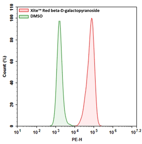
图1.用Xite Red β-D-吡喃半乳糖苷测量β-gal的表达。将9L-LacZ细胞(过度表达β-gal的细胞)与Xite Red β-D-吡喃半乳糖苷在37°C下孵育30分钟。使用NovoCyte流式细胞仪(ACEA Biosciences)通过PE通道获取信号。
|
参考文献
Acridinium Benzoates for Ratiometric Fluorescence Imaging.
Authors: Wen, Min and Wang, Xijing and Wang, Ting and Sun, Yan and Fan, Mengting and Li, Min and Zhu, Junru and Zhang, Dazhi and Cui, Xiaoyan and Shan, Yongkui
Journal: Chemistry (Weinheim an der Bergstrasse, Germany) (2020): 3247-3251
Fluorescence Signal Amplification by Using β-Galactosidase for Flow Cytometry; Advantages of an Endogenous Activity-Free Enzyme.
Authors: Nobori, Takanobu and Kawamura, Masumi and Yoshida, Ryosuke and Joichi, Taisei and Kamino, Kenta and Kishimura, Akihiro and Baba, Eishi and Mori, Takeshi and Katayama, Yoshiki
Journal: Analytical chemistry (2020): 3069-3076
Amphiphilic triphenylamine-benzothiadiazole dyes: preparation, fluorescence and aggregation behavior, and enzyme fluorescence detection.
Authors: Ishi-I, Tsutomu and Kawai, Kazuki and Shirai, Yuya and Kitahara, Ikumi and Hagiwara, Yoshinori
Journal: Photochemical & photobiological sciences : Official journal of the European Photochemistry Association and the European Society for Photobiology (2019): 1447-1460
Macrotheranostic Probe with Disease-Activated Near-Infrared Fluorescence, Photoacoustic, and Photothermal Signals for Imaging-Guided Therapy.
Authors: Zhen, Xu and Zhang, Jianjian and Huang, Jiaguo and Xie, Chen and Miao, Qingqing and Pu, Kanyi
Journal: Angewandte Chemie (International ed. in English) (2018): 7804-7808
Impact of plasma protein binding on cargo release by thermosensitive liposomes probed by fluorescence correlation spectroscopy.
Authors: Mittag, Judith J and Kneidl, Barbara and Preiβ, Tobias and Hossann, Martin and Winter, Gerhard and Wuttke, Stefan and Engelke, Hanna and Rädler, Joachim O
Journal: European journal of pharmaceutics and biopharmaceutics : official journal of Arbeitsgemeinschaft fur Pharmazeutische Verfahrenstechnik e.V (2017): 215-223
Isolation and Fluorescence-Activated Cell Sorting of Mouse Keratinocytes Expressing β-Galactosidase.
Authors: Kasper, Maria and Toftgård, Rune and Jaks, Viljar
Journal: Methods in molecular biology (Clifton, N.J.) (2016): 123-36
Small quinolinium-based enzymatic probes via blue-to-red ratiometric fluorescence.
Authors: Wang, Pan and Du, Jiajun and Liu, Huijing and Bi, Guoqiang and Zhang, Guoqing
Journal: The Analyst (2016): 1483-7
Sensitive β-galactosidase-targeting fluorescence probe for visualizing small peritoneal metastatic tumours in vivo.
Authors: Asanuma, Daisuke and Sakabe, Masayo and Kamiya, Mako and Yamamoto, Kyoko and Hiratake, Jun and Ogawa, Mikako and Kosaka, Nobuyuki and Choyke, Peter L and Nagano, Tetsuo and Kobayashi, Hisataka and Urano, Yasuteru
Journal: Nature communications (2015): 6463
A fluorescence-based method coupled with Disruptor filtration for rapid detection of F + RNA phages.
Authors: Yang, Y and Griffiths, M W
Journal: Letters in applied microbiology (2014): 177-83
Quantitative Fluorescence Assays Using a Self-Powered Paper-Based Microfluidic Device and a Camera-Equipped Cellular Phone.
Authors: Thom, Nicole K and Lewis, Gregory G and Yeung, Kimy and Phillips, Scott T
Journal: RSC advances (2014): 1334-1340