Texas Red-X,琥珀酰亚胺酯 CAS 199745-67-0 货号474-AAT Bioquest荧光染料

上海金畔生物科技有限公司代理AAT Bioquest荧光染料全线产品,欢迎访问AAT Bioquest荧光染料官网了解更多信息。

Texas Red-X,琥珀酰亚胺酯 CAS 199745-67-0

Texas Red-X,琥珀酰亚胺酯 CAS 199745-67-0

Texas Red-X,琥珀酰亚胺酯 CAS 199745-67-0    货号474 货号 474 存储条件 在零下15度以下保存, 避免光照
规格 5 mg 价格 3924
Ex (nm) 586 Em (nm) 603
分子量 816.94 溶剂 DMSO
产品详细介绍

简要概述

产品基本信息

货号:474

产品名称:Texas Red-X,琥珀酰亚胺酯

CAS:199745-67-0

规格:5mg

储存条件:-15℃,避光干燥

保质期:12个月


物理化学光谱特性

分子量:816.94

溶剂:DMSO

Ex(nm):585

Em(nm):602


产品介绍

Texas Red-X,琥珀酰亚胺酯是美国AAT Bioquest生产的荧光染料,Texas Red-X,琥珀酰亚胺酯(胺反应性德克萨斯红®-X,琥珀酰亚胺酯)是Texas Red的优良替代品。 它具有与Texas Red相同的光谱特性,但它更容易与生物膜结合,具有更高的共轭产率。 它可用于产生具有激发/发射最大值~595 / 615nm的亮红色荧光生物共轭物。与TexasRed®相比,该活性染料在荧光团和琥珀酰亚胺酯基团之间含有另外的七个原子氨基己酰基间隔基(“X”)。 该间隔基有助于将荧光团与其连接点分离,潜在地减少荧光团与其缀合的生物分子的相互作用。金畔生物是AAT Bioquest的中国代理商,为您提供最优质的Texas Red-X,琥珀酰亚胺酯。 

点击查看光谱


参考文献

A comparative study of the potential of solid triglyceride nanostructures coated with chitosan or poly(ethylene glycol) as carriers for oral calcitonin delivery
Authors: Garcia-Fuentes M, Prego C, Torres D, Alonso MJ.
Journal: Eur J Pharm Sci (2005): 133

Bead-based cellular analysis, sorting and multiplexing
Authors: Sanchez-Martin RM, Muzerelle M, Chitkul N, How SE, Mittoo S, Bradley M.
Journal: Chembiochem (2005): 1341

Budding dynamics of multicomponent tubular vesicles
Authors: Li L, Liang X, Lin M, Qiu F, Yang Y.
Journal: J Am Chem Soc (2005): 17996

Emission characteristics of fluorescent labels with respect to temperature changes and subsequent effects on DNA microchip studies
Authors: Liu WT, Wu JH, Li ES, Selamat ES.
Journal: Appl Environ Microbiol (2005): 6453

Fluorescence properties of fluorescein, tetramethylrhodamine and Texas Red linked to a DNA aptamer
Authors: Unruh JR, Gokulrangan G, Wilson GS, Johnson CK.
Journal: Photochem Photobiol (2005): 682

Orientational dynamics and dye-DNA interactions in a dye-labeled DNA aptamer
Authors: Unruh JR, Gokulrangan G, Lushington GH, Johnson CK, Wilson GS.
Journal: Biophys J (2005): 3455

Purification and fluorescent labeling of the human serotonin transporter
Authors: Rasmussen SG, Gether U.
Journal: Biochemistry (2005): 3494

Transepithelial transport of fluorescent p-glycoprotein and MRP2 substrates by insect Malpighian tubules: confocal microscopic analysis of secreted fluid droplets
Authors: Leader JP, O’Donnell MJ.
Journal: J Exp Biol (2005): 4363

Measurement of normal and anomalous diffusion of dyes within protein structures fabricated via multiphoton excited cross-linking
Authors: Basu S, Wolgemuth CW, Campagnola PJ.
Journal: Biomacromolecules (2004): 2347

Multistep fluorescence resonance energy transfer in sequential chromophore array constructed on oligo-DNA assemblies
Authors: Ohya Y, Yabuki K, Hashimoto M, Nakajima A, Ouchi T.
Journal: Bioconjug Chem (2003): 1057


相关产品 

产品名称 货号
Texas Red-X,琥珀酰亚胺酯 CAS 216972-99-5 Cat#474

说明书
Texas Red-X,琥珀酰亚胺酯 CAS 199745-67-0.pdf

Ru Red核酸染料 货号TJ1710-AAT Bioquest荧光染料

上海金畔生物科技有限公司代理AAT Bioquest荧光染料全线产品,欢迎访问AAT Bioquest荧光染料官网了解更多信息。

Ru Red核酸染料

Ru Red核酸染料

货号 TJ1710 存储条件 建议在低于-15℃温度下冷冻保存
规格 500 ul 价格 500
Ex (nm) 300 Em (nm) 605
分子量 溶剂 DMSO
产品详细介绍

简要概述

Ru Red是一种结合于所有dsDNA双螺旋小沟区域的具有独特涉及的荧光染料。在游离状态下,Ru Red发出微弱的荧光,但一旦与双链DNA结合后,荧光大大增强。因此,Ru Red的荧光信号强度与双链DNA的数量相关,可以根据荧光信号检测出PCR体系存在的双链DNA数量。Ru Red与 EB 有相同的光谱特性,无需改变滤光片及观察装置。标准的 EB 滤光片或 SYBR 滤光片都适用,使用与观察 EB 相同的普通紫外凝胶透射仪观察即可,在 300 nm 紫外光附近可得到最佳激发。Ru Red的最大吸收波长约为518 nm,发射波长最大约为605 nm。Ru Red 与双链DNA的亲和力非常高,因此可以用做电泳前染色,对分子生物学中常用的酶(如:Taq 酶、逆转录酶、内切酶、T4连接酶等)没有抑制作用。另外,Ru Red 与EB相比,诱变能力大大降低。


Ru Red是高灵敏的DNA荧光染料,适用于各种电泳分析,操作简单:无须脱色或冲洗。至少可检出20pg DNA,高于EB染色法25-100倍。Ru Red 与dsDNA 结合荧光信号会增强800-1000倍,用Ru Red染色的凝胶样品荧光信号强,背景信号低。可适用于多种电泳分析。


Ru Red 适合于多种凝胶电泳方法:琼脂糖凝胶,聚丙烯酰胺凝胶电泳,脉冲电场凝胶电泳,和毛细管电泳等。Ru Red 核酸凝胶染液(Ru Red Nucleic Acid Gel Stains)是一种高敏感性的荧光染液,用于检测琼脂糖凝胶及聚丙烯酰胺凝胶中的双链DNA和寡核苷酸。当染液与核酸结合后,Ru Red 核酸凝胶染液的荧光信号会明显增强,因此经Ru Red 核酸凝胶染液染色的凝胶显示出非常好的信躁比,没有背景荧光。为获得最佳的结果,凝胶最好在跑胶后再进行染色。Ru Red 核酸凝胶染液用于低含量的PCR产物,凋亡研究及异源双链分析中少量DNA的检测较为理想。可应用于:DNA和RNA的检测;SSCP及异源双链分析。

产品说明书

实验方案

1.Ru Red预染方法

1.1该方法适用于琼脂糖凝胶电泳

1.2按照实验要求配制一定浓度的琼脂糖凝胶溶液并用微波炉或其他工具加热熔解。

1.3待加热后的琼脂糖凝胶溶液稍凉(约50-70℃),按1:10000比例加入Ru Red 原液摇匀(使Ru Red工作浓度为1 x)。

1.4倒胶固定,使自然凝固。

1.5加样、电泳,电泳程序根据实验要求。

1.6凝胶观测,成像分析。观测波长与EB相同,也可用SYBR or GelStar等滤光片成像,效果相同。


2.Ru Red后染方法

2.1按照常规方法进行电泳。

2.2用PH 7.0 – 8.5 的缓冲液(如:TAE,TBE 或 TE),按照3300:1的比例稀释Ru Red浓缩液,混匀,制成3X染色溶液。

2.3将3X染色溶液倒入合适的聚丙烯容器中,放入凝胶,用铝箔等盖住容器使染料避光。室温振荡染色10-30分钟,染色时间因凝胶浓度和厚度而定。聚丙烯酰胺凝胶直接在玻璃平皿上染色,将配好的工作溶液轻轻地倒在胶板上,让工作液均匀地覆盖整个胶板,并染色30分钟。玻璃平皿必须预先经过硅烷化溶液处理(避免染料吸附在玻璃表面上)。

2.4使用蓝光透照仪观测。蓝光可透过玻璃,观测聚丙烯酰胺凝胶时,可直接将托有凝胶的玻璃平皿放入蓝光透照仪内观测。可使用EB, SYBR 或GelStar记录成像系统。


3.Ru Red使用注意事项

3.1在”Ru Red预染色方法”中,电泳时间不要超过2小时,否则Ru Red会从DNA上分离出来,会产生弥散状条带。

3.2在常规用酒精沉淀核酸的过程中,Ru Red 可以全部从双链核酸上去掉。

3.3如果想对用 Ru Red 染过的胶进行 Southern blots,建议在预杂化和杂化溶液中加入 0.1%- 0.3% 的SDS。

3.4在紫外照射透视下,与双链 DNA 接合的 Ru Red 呈现红色荧光。

3.5Ru Red对玻璃和非聚丙烯材料具有一定亲合力。建议在稀释、贮存、染色等使用过程中用聚丙烯类容器。

钙离子荧光探针Fura Red, AM 货号21046-AAT Bioquest荧光染料

上海金畔生物科技有限公司代理AAT Bioquest荧光染料全线产品,欢迎访问AAT Bioquest荧光染料官网了解更多信息。

钙离子荧光探针Fura Red, AM

钙离子荧光探针Fura Red, AM

钙离子荧光探针Fura Red, AM    货号21046 货号 21046 存储条件 在零下15度以下保存, 避免光照
规格 1 mg 价格 4584
Ex (nm) 435 Em (nm) 639
分子量 1088.99 溶剂 DMSO
产品详细介绍

简要概述

产品基本信息

货号:21046

产品名称:钙离子荧光探针Fura Red, AM

CAS:149732-62-7

规格:1mg

储存条件:-15℃避光防潮

保质期:24个月


产品物理化学光谱特性

分子量:1088.99

溶剂:DMSO

激发波长(nm):458

发射波长(nm):597


适用仪器


荧光酶标仪  
激发: 435,470nm
发射: 630,650nm
cutoff: Ex/Em = 435/630, cutoff 610. Ex/Em = 470/650, cut off 630
推荐孔板: 黑色透明
读取模式: 底读模式


产品介绍

钙离子荧光探针Fura Red, AM是美国AAT Bioquest生产的钙离子荧光探针,Fura Red是一种可见光可激发的fura-2类似物,当与单激发,绿色荧光钙指示剂一起使用时,通过显微光度法,成像或流式细胞术为单细胞中的钙离子比率测量提供了独特的可能性。 Fura Red AM是用于非侵入性细胞内负载的Fura Red的细胞渗透型。 Fura Red AM可以同时装入Fluo-3 AM,Fluo-8 AM或Cal-520 AM的细胞中。 组合两种钙染料的优点是可以使用具有较长激发波长的染料。 与使用用UV或近紫外光(例如Fura-2)激发的比率染料相比,这通常对细胞造成的伤害更小,因为可见光波长的光具有较低的光毒性。金畔生物是AAT Bioquest的中国代理商,为您提供最优质的钙离子荧光探针Fura Red, AM。 

点击查看光谱

点击查看实验方案

钙离子篇:时间轴式讲解应用于钙离子检测的探针


参考文献

Ratiometric analysis of fura red by flow cytometry: a technique for monitoring intracellular calcium flux in primary cell subsets
Authors: Wendt ER, Ferry H, Greaves DR, Keshav S.
Journal: PLoS One (2015): e0119532

A flow cytometric comparison of Indo-1 to fluo-3 and Fura Red excited with low power lasers for detecting Ca(2+) flux
Authors: Bailey S, Macardle PJ.
Journal: J Immunol Methods (2006): 220

Use of co-loaded Fluo-3 and Fura Red fluorescent indicators for studying the cytosolic Ca(2+)concentrations distribution in living plant tissue
Authors: Walczysko P, Wagner E, Albrechtova JT.
Journal: Cell Calcium (2000): 23

[Monitoring calcium in outer hair cells with confocal microscopy and fluorescence ratios of fluo-3 and fura-red]
Authors: Su ZL, Li N, Sun YR, Yang J, Wang IM, Jiang SC.
Journal: Shi Yan Sheng Wu Xue Bao (1998): 323

Calcium transient alternans in blood-perfused ischemic hearts: observations with fluorescent indicator fura red
Authors: Wu Y, Clusin WT.
Journal: Am J Physiol (1997): H2161

Problems associated with using Fura-2 to measure free intracellular calcium concentrations in human red blood cells
Authors: Blackwood AM, Sagnella GA, Markandu ND, MacGregor GA.
Journal: J Hum Hypertens (1997): 601

IgG-induced Ca2+ oscillations in differentiated U937 cells; a study using laser scanning confocal microscopy and co-loaded fluo-3 and fura-red fluorescent probes
Authors: Floto RA, Mahaut-Smith MP, Somasundaram B, Allen JM.
Journal: Cell Calcium (1995): 377

Improved sensitivity in flow cytometric intracellular ionized calcium measurement using fluo-3/Fura Red fluorescence ratios
Authors: Novak EJ, Rabinovitch PS.
Journal: Cytometry (1994): 135

Localization of calcium entry through calcium channels in olfactory receptor neurones using a laser scanning microscope and the calcium indicator dyes Fluo-3 and Fura-Red
Authors: Schild D, Jung A, Schultens HA.
Journal: Cell Calcium (1994): 341

The distribution of intracellular calcium chelator (fura-2) in a population of intact human red cells
Authors: Lew VL, Etzion Z, Bookchin RM, daCosta R, Vaananen H, Sassaroli M, Eisinger J.
Journal: Biochim Biophys Acta (1993): 152

说明书
钙离子荧光探针Fura Red, AM.pdf

Texas Red-X,琥珀酰亚胺酯(混合异构体) CAS 216972-99-5 货号475-AAT Bioquest荧光染料

上海金畔生物科技有限公司代理AAT Bioquest荧光染料全线产品,欢迎访问AAT Bioquest荧光染料官网了解更多信息。

Texas Red-X,琥珀酰亚胺酯(混合异构体) CAS 216972-99-5

Texas Red-X,琥珀酰亚胺酯(混合异构体) CAS 216972-99-5

Texas Red-X,琥珀酰亚胺酯(混合异构体) CAS 216972-99-5    货号475 货号 475 存储条件 在零下15度以下保存, 避免光照
规格 5 mg 价格 2604
Ex (nm) 586 Em (nm) 603
分子量 816.94 溶剂 DMSO
产品详细介绍

简要概述

产品基本信息

货号:475

产品名称:Texas Red-X,琥珀酰亚胺酯(混合异构体)

CAS:216972-99-5

规格:5mg

储存条件:-15℃,避光干燥

保质期:12个月


物理化学光谱特性

分子量:816.94

溶剂:DMSO

Ex(nm):585

Em(nm):602


产品介绍

Texas Red-X,琥珀酰亚胺酯是美国AAT Bioquest生产的荧光染料,Texas Red-X,琥珀酰亚胺酯(胺反应性德克萨斯红®-X,琥珀酰亚胺酯)是Texas Red的优良替代品。 它具有与Texas Red相同的光谱特性,但它更容易与生物膜结合,具有更高的共轭产率。 它可用于产生具有激发/发射最大值~595 / 615nm的亮红色荧光生物共轭物。与TexasRed®相比,该活性染料在荧光团和琥珀酰亚胺酯基团之间含有另外的七个原子氨基己酰基间隔基(“X”)。 该间隔基有助于将荧光团与其连接点分离,潜在地减少荧光团与其缀合的生物分子的相互作用。金畔生物是AAT Bioquest的中国代理商,为您提供最优质的Texas Red-X,琥珀酰亚胺酯。 

点击查看光谱


参考文献

A comparative study of the potential of solid triglyceride nanostructures coated with chitosan or poly(ethylene glycol) as carriers for oral calcitonin delivery
Authors: Garcia-Fuentes M, Prego C, Torres D, Alonso MJ.
Journal: Eur J Pharm Sci (2005): 133

Bead-based cellular analysis, sorting and multiplexing
Authors: Sanchez-Martin RM, Muzerelle M, Chitkul N, How SE, Mittoo S, Bradley M.
Journal: Chembiochem (2005): 1341

Budding dynamics of multicomponent tubular vesicles
Authors: Li L, Liang X, Lin M, Qiu F, Yang Y.
Journal: J Am Chem Soc (2005): 17996

Emission characteristics of fluorescent labels with respect to temperature changes and subsequent effects on DNA microchip studies
Authors: Liu WT, Wu JH, Li ES, Selamat ES.
Journal: Appl Environ Microbiol (2005): 6453

Fluorescence properties of fluorescein, tetramethylrhodamine and Texas Red linked to a DNA aptamer
Authors: Unruh JR, Gokulrangan G, Wilson GS, Johnson CK.
Journal: Photochem Photobiol (2005): 682

Orientational dynamics and dye-DNA interactions in a dye-labeled DNA aptamer
Authors: Unruh JR, Gokulrangan G, Lushington GH, Johnson CK, Wilson GS.
Journal: Biophys J (2005): 3455

Purification and fluorescent labeling of the human serotonin transporter
Authors: Rasmussen SG, Gether U.
Journal: Biochemistry (2005): 3494

Transepithelial transport of fluorescent p-glycoprotein and MRP2 substrates by insect Malpighian tubules: confocal microscopic analysis of secreted fluid droplets
Authors: Leader JP, O’Donnell MJ.
Journal: J Exp Biol (2005): 4363

Measurement of normal and anomalous diffusion of dyes within protein structures fabricated via multiphoton excited cross-linking
Authors: Basu S, Wolgemuth CW, Campagnola PJ.
Journal: Biomacromolecules (2004): 2347

Multistep fluorescence resonance energy transfer in sequential chromophore array constructed on oligo-DNA assemblies
Authors: Ohya Y, Yabuki K, Hashimoto M, Nakajima A, Ouchi T.
Journal: Bioconjug Chem (2003): 1057


相关产品

产品名称 货号
Texas Red-X,琥珀酰亚胺酯 Cat#475

说明书
Texas Red-X,琥珀酰亚胺酯(混合异构体) CAS 216972-99-5.pdf

Xite Red β-D-吡喃半乳糖苷*红色荧光* 货号14035-AAT Bioquest荧光染料

上海金畔生物科技有限公司代理AAT Bioquest荧光染料全线产品,欢迎访问AAT Bioquest荧光染料官网了解更多信息。

Xite Red β-D-吡喃半乳糖苷*红色荧光*

Xite Red β-D-吡喃半乳糖苷*红色荧光*

Xite Red β-D-吡喃半乳糖苷*红色荧光*    货号14035 货号 14035 存储条件 在零下15度以下保存, 避免光照
规格 1 mg 价格 2472
Ex (nm) Em (nm)
分子量 591.63 溶剂 DMSO
产品详细介绍

简要概述

Xite Red β-D-半乳糖吡喃糖苷是β-半乳糖苷酶(β-gal)的荧光底物。与现有的β-半乳糖苷酶底物(例如,常用的FDG)相比,它具有更好的细胞通透性。Xite Red β-D-吡喃半乳糖苷很容易进入细胞,并被β-gal裂解,从而产生强荧光产品Xite Red。强烈荧光的Xite Red可以很好地保留在细胞中,从而易于通过流式细胞仪和荧光显微镜进行检测。Xite Red β-D-半乳糖吡喃糖苷提供了一种简单而灵敏的工具来检测β-半乳糖苷酶的活性。Xite Red β-D-吡喃半乳糖苷可以作为检测细胞中细胞衰老的工具,因为β-gal已被确定为细胞衰老的可靠标记。此外,Xite Red β-D-吡喃半乳糖苷是可固定的。Xite Red β-D-吡喃半乳糖苷产生的红色荧光可以很容易地与其他颜色的荧光探针(例如DAPI或GFP)结合使用,以进行多色荧光分析。金畔生物是AAT Bioquest的中国代理商,为您提供最优质的Xite Red β-D-半乳糖吡喃糖苷。 


适用仪器


流式细胞仪  
激发: 488nm激光
发射: 575/26 nm滤波片
通道: PE滤波片组
荧光显微镜  
激发: Cy3/TRITC滤波片
发射: Cy3/TRITC滤波片
推荐孔板: 黑色透明


产品说明书

样品实验方案

以下是我们推荐的方案,仅提供指导。具体实验应根据您的特定需求进行修改。

简要概述

  1. 根据需要处理样品
  2. 准备Xite Red β-D-吡喃半乳糖苷工作溶液并将其添加到样品中
  3. 在37°C下孵育样品15至45分钟
  4. 使用带有575/26 nm滤光片的流式细胞仪(PE通道)或带有Cy3/TRITC滤光片组的荧光显微镜检测荧光强度 


溶液配制

储备溶液配制

Xite Red β-D-吡喃半乳糖苷储备溶液:在Xite Red β-D-吡喃半乳糖苷中加入适量的DMSO,制成2-5 mM Xite Red β-D-吡喃半乳糖苷原液。注意:将未使用的Xite Red β D-吡喃半乳糖苷原液以等份储存在-20°C下。
 
工作溶液配制
Xite Red β-D-吡喃半乳糖苷工作溶液:在自备的缓冲液中制备1-20 µM Xite Red β-D-吡喃半乳糖苷工作溶液。注意1:Xite Red β-D-吡喃半乳糖苷工作溶液应立即使用。注意2:Xite Red β-D-吡喃半乳糖苷的浓度应针对不同的细胞类型和条件进行优化。


操作步骤

  1. 根据需要处理样品。
  2. 处理并用自备的缓冲液(例如DPBS)洗涤细胞。
  3. 加入Xite Red β-D-吡喃半乳糖苷工作溶液15-45分钟,然后在37°C的培养箱中培养样品。
    注意:孵育的最佳时间需要通过实验确定。
  4. 取出工作溶液并用自备的缓冲液洗涤细胞。
  5. 将细胞重悬在自备的缓冲液中,并使用流式细胞仪使用575/26 nm滤光片(PE通道)或带有Cy3/TRITC滤光片组的荧光显微镜检测荧光强度。


图示


Xite Red β-D-吡喃半乳糖苷*红色荧光*    货号14035

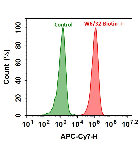
图1.用Xite Red β-D-吡喃半乳糖苷测量β-gal的表达。将9L-LacZ细胞(过度表达β-gal的细胞)与Xite Red β-D-吡喃半乳糖苷在37°C下孵育30分钟。使用NovoCyte流式细胞仪(ACEA Biosciences)通过PE通道获取信号。


参考文献

Acridinium Benzoates for Ratiometric Fluorescence Imaging.
Authors: Wen, Min and Wang, Xijing and Wang, Ting and Sun, Yan and Fan, Mengting and Li, Min and Zhu, Junru and Zhang, Dazhi and Cui, Xiaoyan and Shan, Yongkui
Journal: Chemistry (Weinheim an der Bergstrasse, Germany) (2020): 3247-3251

Fluorescence Signal Amplification by Using β-Galactosidase for Flow Cytometry; Advantages of an Endogenous Activity-Free Enzyme.
Authors: Nobori, Takanobu and Kawamura, Masumi and Yoshida, Ryosuke and Joichi, Taisei and Kamino, Kenta and Kishimura, Akihiro and Baba, Eishi and Mori, Takeshi and Katayama, Yoshiki
Journal: Analytical chemistry (2020): 3069-3076

Amphiphilic triphenylamine-benzothiadiazole dyes: preparation, fluorescence and aggregation behavior, and enzyme fluorescence detection.
Authors: Ishi-I, Tsutomu and Kawai, Kazuki and Shirai, Yuya and Kitahara, Ikumi and Hagiwara, Yoshinori
Journal: Photochemical & photobiological sciences : Official journal of the European Photochemistry Association and the European Society for Photobiology (2019): 1447-1460

Macrotheranostic Probe with Disease-Activated Near-Infrared Fluorescence, Photoacoustic, and Photothermal Signals for Imaging-Guided Therapy.
Authors: Zhen, Xu and Zhang, Jianjian and Huang, Jiaguo and Xie, Chen and Miao, Qingqing and Pu, Kanyi
Journal: Angewandte Chemie (International ed. in English) (2018): 7804-7808

Impact of plasma protein binding on cargo release by thermosensitive liposomes probed by fluorescence correlation spectroscopy.
Authors: Mittag, Judith J and Kneidl, Barbara and Preiβ, Tobias and Hossann, Martin and Winter, Gerhard and Wuttke, Stefan and Engelke, Hanna and Rädler, Joachim O
Journal: European journal of pharmaceutics and biopharmaceutics : official journal of Arbeitsgemeinschaft fur Pharmazeutische Verfahrenstechnik e.V (2017): 215-223

Isolation and Fluorescence-Activated Cell Sorting of Mouse Keratinocytes Expressing β-Galactosidase.
Authors: Kasper, Maria and Toftgård, Rune and Jaks, Viljar
Journal: Methods in molecular biology (Clifton, N.J.) (2016): 123-36

Small quinolinium-based enzymatic probes via blue-to-red ratiometric fluorescence.
Authors: Wang, Pan and Du, Jiajun and Liu, Huijing and Bi, Guoqiang and Zhang, Guoqing
Journal: The Analyst (2016): 1483-7

Sensitive β-galactosidase-targeting fluorescence probe for visualizing small peritoneal metastatic tumours in vivo.
Authors: Asanuma, Daisuke and Sakabe, Masayo and Kamiya, Mako and Yamamoto, Kyoko and Hiratake, Jun and Ogawa, Mikako and Kosaka, Nobuyuki and Choyke, Peter L and Nagano, Tetsuo and Kobayashi, Hisataka and Urano, Yasuteru
Journal: Nature communications (2015): 6463

A fluorescence-based method coupled with Disruptor filtration for rapid detection of F + RNA phages.
Authors: Yang, Y and Griffiths, M W
Journal: Letters in applied microbiology (2014): 177-83

Quantitative Fluorescence Assays Using a Self-Powered Paper-Based Microfluidic Device and a Camera-Equipped Cellular Phone.
Authors: Thom, Nicole K and Lewis, Gregory G and Yeung, Kimy and Phillips, Scott T
Journal: RSC advances (2014): 1334-1340

说明书
Xite Red β-D-吡喃半乳糖苷*红色荧光*.pdf