核绿 DCS1 死细胞染料 货号17550-AAT Bioquest荧光染料

上海金畔生物科技有限公司代理AAT Bioquest荧光染料全线产品,欢迎访问AAT Bioquest荧光染料官网了解更多信息。

核绿 DCS1 死细胞染料

核绿 DCS1 死细胞染料

核绿 DCS1 死细胞染料    货号17550 货号 17550 存储条件 在零下15度以下保存, 避免光照
规格 0.5 ml 价格 1272
Ex (nm) 503 Em (nm) 527
分子量 N/A 溶剂 DMSO
产品详细介绍

简要概述

产品基本信息

货号:17550

产品名称:核绿 DCS1 死细胞染料

规格:0.5ml

储存条件:-20℃干燥


产品物理化学光谱特性

溶剂:DMSO

激发波长(nm):503

发射波长(nm):526


产品介绍

我们的Nuclear Green DCS1是一种发荧光的,DNA选择性和不渗透细胞的染料,用于分析死亡,固定或凋亡细胞中的DNA含量。当与DNA结合后,Nuclear Green DCS1的绿色荧光显着增强。它可用于荧光成像,微孔板和流式细胞仪应用。这种具有DNA结合力的染料可用于通过适当过滤器设置对死亡,固定或凋亡细胞进行多色分析。

点击查看光谱

产品说明书

染色细胞分析方案

注意:以下协议适用于大多数细胞类型。 生长培养基,细胞密度,其他细胞类型和因子的存在可能影响染色。 玻璃器皿上的残留洗涤剂也可能影响许多生物的染色,并导致在有或没有细胞存在的溶液中出现明亮染色的物质。

1.将Nuclear Green DCS1(2至10μM)加入固定的,死亡的或凋亡的细胞(悬浮液或粘附细胞)中,并将细胞染色15至60分钟。 在最初的实验中,最好尝试几种染料浓度来确定产生所需结果的最佳浓度。 高染料浓度倾向于引起其他细胞结构的非特异性染色。

2.用荧光显微镜,荧光酶标仪或流式细胞仪直接分析细胞染色。


参考文献

One-Step Seeding of Neural Stem Cells with Vitronectin-Supplemented Medium for High-Throughput Screening Assays
Authors: Sheng Dai, Rong Li, Yan Long, Steve Titus, Jinghua Zhao, Ruili Huang, Menghang Xia, Wei Zheng
Journal: Journal of Biomolecular Screening (2016): 1112–1124

Genome shuffling of the nonconventional yeast Pichia anomala for improved sugar alcohol production
Authors: Guoqiang Zhang, Yuping Lin, Xianni Qi, Lixian Wang, Peng He, Qinhong Wang, Yanhe Ma
Journal: Microbial cell factories (2015): 1


相关产品

产品名称 货号
核绿 LCS1 活细胞染料 Cat#17540
核红 LCS1 活细胞染料 Cat#17542
核紫 LCS1 活细胞染料 Cat#17543

说明书
核绿 DCS1 死细胞染料.pdf

核红 DCS1 死细胞染料 货号17552-AAT Bioquest荧光染料

上海金畔生物科技有限公司代理AAT Bioquest荧光染料全线产品,欢迎访问AAT Bioquest荧光染料官网了解更多信息。

核红 DCS1 死细胞染料

核红 DCS1 死细胞染料

核红 DCS1 死细胞染料    货号17552 货号 17552 存储条件 在零下15度以下保存, 避免光照
规格 0.5 ml 价格 1272
Ex (nm) 631 Em (nm) 651
分子量 671.42 溶剂 DMSO
产品详细介绍

简要概述

产品基本信息

货号:17552

产品名称:核红 DCS1 死细胞染料

规格:0.5ml

储存条件:-20℃干燥


产品物理化学光谱特性

溶剂:DMSO

激发波长(nm):642

发射波长(nm):660


产品介绍

我们的Nuclear Red DCS1是一种发荧光的,DNA选择性和不渗透细胞的染料,用于分析死细胞,固定细胞或凋亡细胞中的DNA含量。结合DNA时,Nuclear Red DCS1的红色荧光显着增强。它可用于荧光成像,微孔板和流式细胞仪应用。这种具有DNA结合力的染料可用于通过适当过滤器设置对死亡,固定或凋亡细胞进行多色分析。

点击查看光谱

产品说明书

染色细胞分析方案

注意:以下协议适用于大多数细胞类型。 生长培养基,细胞密度,其他细胞类型和因子的存在可能影响染色。 玻璃器皿上的残留洗涤剂也可能影响许多生物的染色,并导致在有或没有细胞存在的溶液中出现明亮染色的物质。

1.将Nuclear Red DCS1(2至10μM)加入固定的,死亡或凋亡的细胞(悬浮液或粘附细胞)中,并将细胞染色15至60分钟。 在最初的实验中,最好尝试几种染料浓度来确定产生所需结果的最佳浓度。 高染料浓度倾向于引起其他细胞结构的非特异性染色。

2.用荧光显微镜,荧光酶标仪或流式细胞仪直接分析细胞染色。


试剂应用文献

Neural stem cells for disease modeling and evaluation of therapeutics for infantile (CLN1/PPT1) and late infantile (CLN2/TPP1) neuronal ceroid lipofuscinoses


参考文献

Neural stem cells for disease modeling and evaluation of therapeutics for infantile (CLN1/PPT1) and late infantile (CLN2/TPP1) neuronal ceroid lipofuscinoses
Authors: Ni Sima, Rong Li, Wei Huang, Miao Xu, Jeanette Beers, Jizhong Zou, Steven Titus, Elizabeth A Ottinger, Juan J Marugan, Xing Xie
Journal: Orphanet journal of rare diseases (2018): 54

Synthesis of the Anionic Hydroxypropyl-β-cyclodextrin: Poly (decamethylenephosphate) Polyrotaxane and Evaluation of its Cholesterol Efflux Potential in Niemann-Pick C1 Cells
Authors: Kerstin Egele, Shayak Samaddar, Nina Schneider, David H Thompson, Gerhard Wenz
Journal: Journal of Materials Chemistry B (2018)

One-Step Seeding of Neural Stem Cells with Vitronectin-Supplemented Medium for High-Throughput Screening Assays
Authors: Sheng Dai, Rong Li, Yan Long, Steve Titus, Jinghua Zhao, Ruili Huang, Menghang Xia, Wei Zheng
Journal: Journal of Biomolecular Screening (2016): 1112–1124

Genome shuffling of the nonconventional yeast Pichia anomala for improved sugar alcohol production
Authors: Guoqiang Zhang, Yuping Lin, Xianni Qi, Lixian Wang, Peng He, Qinhong Wang, Yanhe Ma
Journal: Microbial cell factories (2015): 1


相关产品

产品名称 货号
核绿 LCS1 活细胞染料 Cat#17540
核红 LCS1 活细胞染料 Cat#17542
核紫 LCS1 活细胞染料 Cat#17543

说明书
核红 DCS1 死细胞染料.pdf

核橙 DCS1 死细胞染料 货号17551-AAT Bioquest荧光染料

上海金畔生物科技有限公司代理AAT Bioquest荧光染料全线产品,欢迎访问AAT Bioquest荧光染料官网了解更多信息。

核橙 DCS1 死细胞染料

核橙 DCS1 死细胞染料

核橙 DCS1 死细胞染料    货号17551 货号 17551 存储条件 在零下15度以下保存, 避免光照
规格 0.5 ml 价格 1272
Ex (nm) 514 Em (nm) 556
分子量 N/A 溶剂 DMSO
产品详细介绍

简要概述

产品基本信息

货号:17551

产品名称:核橙 DCS1 死细胞染料

规格:0.5 ml

储存条件:-20℃干燥


产品物理化学光谱特性

溶剂:DMSO

激发波长(nm):528

发射波长(nm):576


产品介绍

我们的Nuclear Orange DCS1是一种荧光染料,DNA选择性和不渗透细胞的染料,可用于分析死亡,固定或凋亡细胞中的DNA含量。在与DNA结合后,Nuclear Orange DCS1的橙色荧光显着增强。它可用于荧光成像,微孔板和流式细胞仪应用。这种具有DNA结合力的染料可用于通过适当过滤器设置对死亡,固定或凋亡细胞进行多色分析。

点击查看光谱

产品说明书

染色细胞分析方案

注意:以下协议适用于大多数细胞类型。 生长培养基,细胞密度,其他细胞类型和因子的存在可能影响染色。 玻璃器皿上的残留洗涤剂也可能影响许多生物的染色,并导致在有或没有细胞存在的溶液中出现明亮染色的物质。

1.将Nuclear Green DCS1(2至10μM)加入固定的,死亡的或凋亡的细胞(悬浮液或粘附细胞)中,并将细胞染色15至60分钟。 在最初的实验中,最好尝试几种染料浓度来确定产生所需结果的最佳浓度。 高染料浓度倾向于引起其他细胞结构的非特异性染色。

2.用荧光显微镜,荧光酶标仪或流式细胞仪直接分析细胞染色。


参考文献

One-Step Seeding of Neural Stem Cells with Vitronectin-Supplemented Medium for High-Throughput Screening Assays
Authors: Sheng Dai, Rong Li, Yan Long, Steve Titus, Jinghua Zhao, Ruili Huang, Menghang Xia, Wei Zheng
Journal: Journal of Biomolecular Screening (2016): 1112–1124

Genome shuffling of the nonconventional yeast Pichia anomala for improved sugar alcohol production
Authors: Guoqiang Zhang, Yuping Lin, Xianni Qi, Lixian Wang, Peng He, Qinhong Wang, Yanhe Ma
Journal: Microbial cell factories (2015): 1


相关产品

产品名称 货号
核绿 LCS1 活细胞染料 Cat#17540
核红 LCS1 活细胞染料 Cat#17542
核紫 LCS1 活细胞染料 Cat#17543

说明书
核橙 DCS1 死细胞染料.pdf

核紫 DCS1 死细胞染料 货号17549-AAT Bioquest荧光染料

上海金畔生物科技有限公司代理AAT Bioquest荧光染料全线产品,欢迎访问AAT Bioquest荧光染料官网了解更多信息。

核紫 DCS1 死细胞染料

核紫 DCS1 死细胞染料

核紫 DCS1 死细胞染料    货号17549 货号 17549 存储条件 在零下15度以下保存, 避免光照
规格 0.5 ml 价格 1164
Ex (nm) 371 Em (nm) 454
分子量 744.77 溶剂 DMSO
产品详细介绍

简要概述

产品基本信息

货号:17549

产品名称:核紫 DCS1 死细胞染料

规格:0.5ml

储存条件:-20℃干燥


产品物理化学光谱特性

溶剂:DMSO

激发波长(nm):371

发射波长(nm):454


产品介绍

我们的Nuclear Violet DCS1是一种非荧光,DNA选择性和细胞不透性的紫色荧光染料,用于分析死细胞中的DNA含量。 核紫色DCS1在与双链DNA结合后具有明显增强的荧光。 它可用于荧光成像,微孔板和流式细胞仪应用。 这种DNA结合染料可用于死细胞的多色分析。

点击查看光谱


适用仪器


荧光显微镜  
激发: DAPI
发射: DAPI
推荐孔板: 黑色透明孔板

产品说明书

染色细胞分析方案

注意:以下方案适用于大多数细胞类型。 生长培养基,细胞密度,其他细胞类型和因子的存在可能影响染色。 玻璃器皿上的残留洗涤剂也可能影响许多生物的染色,并导致在有或没有细胞存在的溶液中出现明亮染色的物质。


工作溶液配制

使用缓冲液将核紫 DCS1储备溶液(5mM)稀释到0.5-5μm的浓度。


操作步骤

1.将Nuclear Violet DCS1工作溶液加到固定、死亡或凋亡的细胞(悬浮液或粘附细胞)中,并将细胞染色15至60分钟。 在最初的实验中,最好尝试几种染料浓度来确定产生所需结果的最佳浓度。 高染料浓度倾向于引起其他细胞结构的非特异性染色。

2.用荧光显微镜、荧光酶标仪或流式细胞仪直接检测染色细胞。
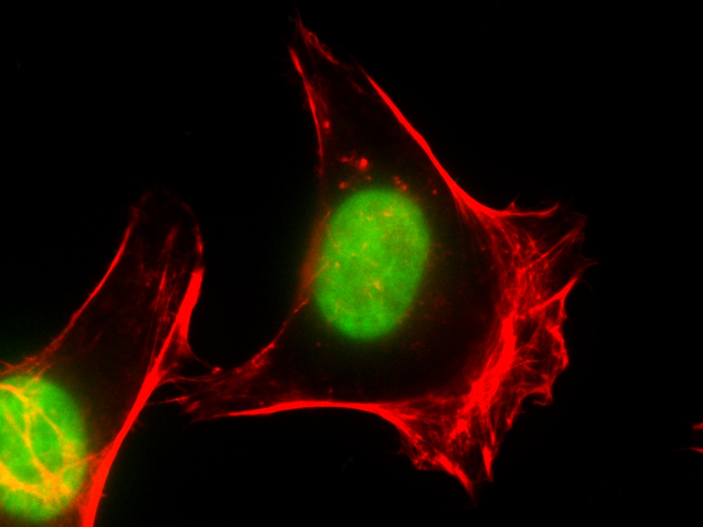
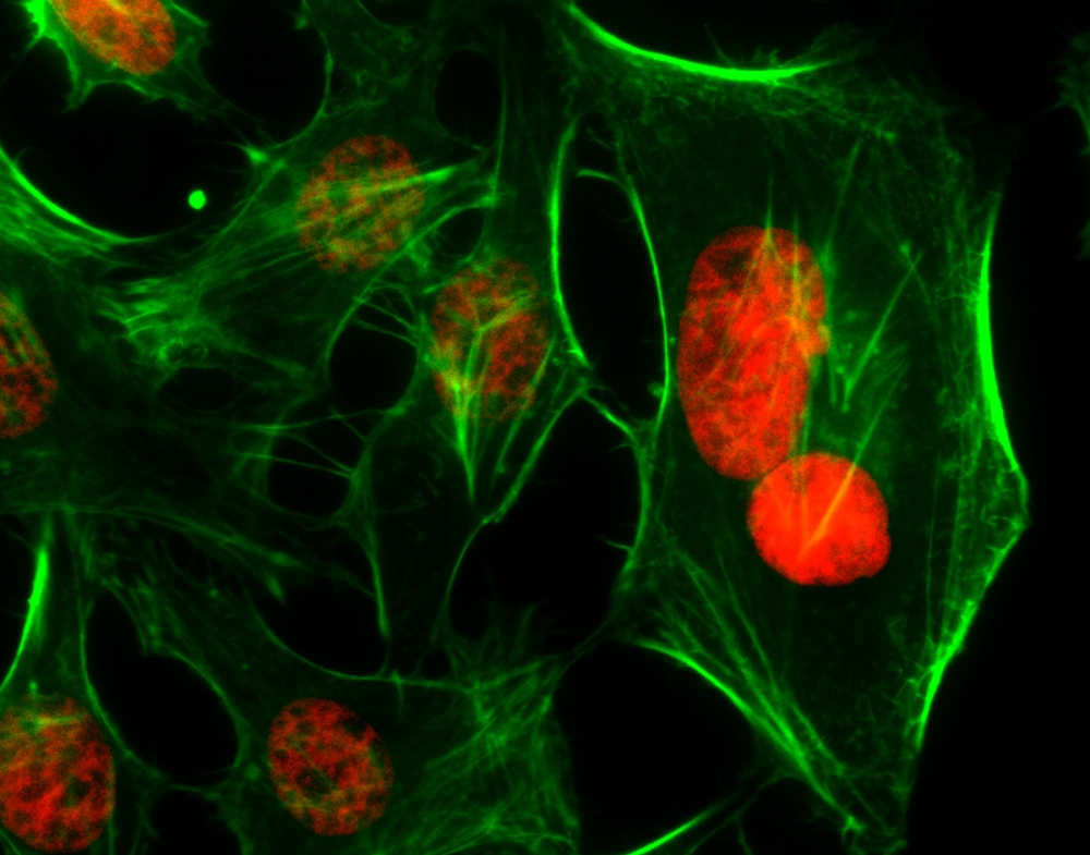
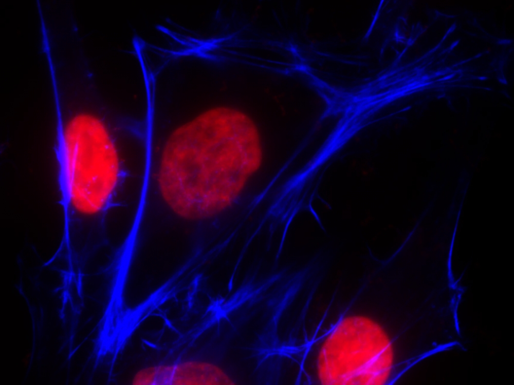
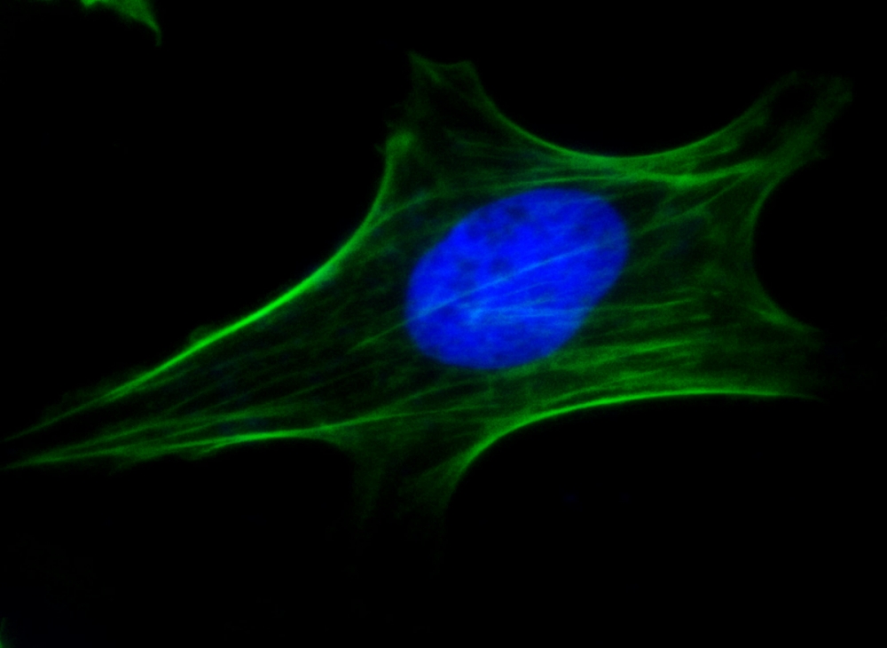
 

图示

核紫 DCS1 死细胞染料    货号17549

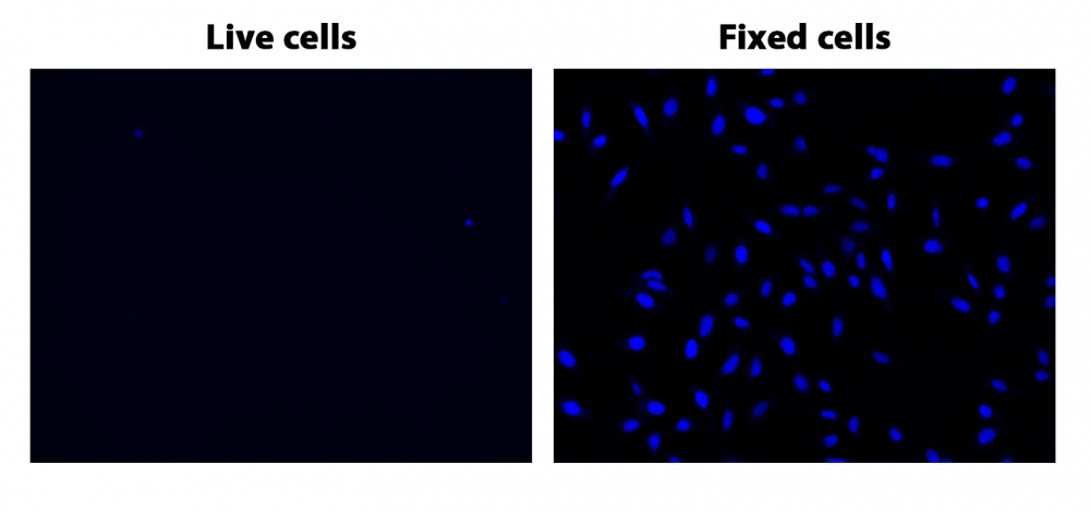
图1.在96孔板上染色的固定和活(非固定)Hela细胞,与核蓝 DCS1 1μm孵育20分钟并在DAPI通道成像。


参考文献

Assessment of laser-induced thermal damage in fresh skin with ex vivo confocal microscopy.
Authors: Ortner, Vinzent Kevin and Sahu, Aditi and Haedersdal, Merete and Rajadhyaksha, Milind and Rossi, Anthony Mario
Journal: Journal of the American Academy of Dermatology (2021): e19-e21

Development of a novel flow cytometry-based approach for reticulocytes micronucleus test in rat peripheral blood.
Authors: Chen, Yiyi and Huo, Jiao and Liu, Yunjie and Zeng, Zhu and Zhu, Xuejiao and Chen, Xuxi and Wu, Rui and Zhang, Lishi and Chen, Jinyao
Journal: Journal of applied toxicology : JAT (2021): 595-606

Exploring the utility of Deep Red Anthraquinone 5 for digital staining of ex vivo confocal micrographs of optically sectioned skin.
Authors: Ortner, Vinzent Kevin and Sahu, Aditi and Cordova, Miguel and Kose, Kivanc and Aleissa, Saud and Alessi-Fox, Christi and Haedersdal, Merete and Rajadhyaksha, Milind and Rossi, Anthony Mario
Journal: Journal of biophotonics (2021): e202000207

A novel method to purify neutrophils enables functional analysis of zebrafish hematopoiesis.
Authors: Konno, Katsuhiro and Kulkeaw, Kasem and Sasada, Manabu and Nii, Takenobu and Kaneyuki, Ayako and Ishitani, Tohru and Arai, Fumio and Sugiyama, Daisuke
Journal: Genes to cells : devoted to molecular & cellular mechanisms (2020): 770-781

Analysis of erythroid maturation in the nonlysed bone marrow with help of radar plots facilitates detection of flow cytometric aberrations in myelodysplastic syndromes.
Authors: Violidaki, Despoina and Axler, Olof and Jafari, Katayoon and Bild, Filippa and Nilsson, Lars and Mazur, Joanna and Ehinger, Mats and Porwit, Anna
Journal: Cytometry. Part B, Clinical cytometry (2020): 399-411

Identification of Cancer-Associated Circulating Cells in Anal Cancer Patients.
Authors: Carter, Thomas J and Jeyaneethi, Jeyarooban and Kumar, Juhi and Karteris, Emmanouil and Glynne-Jones, Rob and Hall, Marcia
Journal: Cancers (2020)

Influence of Polymer Charge on the Localization and Dark- and Photo-Induced Toxicity of a Potential Type I Photosensitizer in Cancer Cell Models.
Authors: Lindgren, Mikael and Gederaas, Odrun A and Siksjø, Monica and Hansen, Tom A and Chen, Lena and Mettra, Bastien and Andraud, Chantal and Monnereau, Cyrille
Journal: Molecules (Basel, Switzerland) (2020)

TEM observation of compacted DNA of Synechococcus elongatus PCC 7942 using DRAQ5 labelling with DAB photooxidation and osmium black.
Authors: Ghosh, Ilika and Atsuzawa, Kimie and Arai, Aoi and Ohmukai, Ryuzo and Kaneko, Yasuko
Journal: Microscopy (Oxford, England) (2020)

[The Establishment of a Three-color Flow Cytometry Approach for the Scoring of Micronucleated Reticulocytes in Rat Bone Marrow].
Authors: Zeng, Zhu and Zhu, Xue-Jiao and Huo, Jiao and Liu, Yun-Jie and Peng, Zi-Hao and Chen, Jin-Yao and Zhang, Li-Shi
Journal: Sichuan da xue xue bao. Yi xue ban = Journal of Sichuan University. Medical science edition (2020): 67-73

Automated Triage Radiation Biodosimetry: Integrating Imaging Flow Cytometry with High-Throughput Robotics to Perform the Cytokinesis-Block Micronucleus Assay.
Authors: Wang, Qi and Rodrigues, Matthew A and Repin, Mikhail and Pampou, Sergey and Beaton-Green, Lindsay A and Perrier, Jay and Garty, Guy and Brenner, David J and Turner, Helen C and Wilkins, Ruth C
Journal: Radiation research (2019): 342-351


相关产品

产品名称 货号
核绿 LCS1 活细胞染料 Cat#17540
核红 LCS1 活细胞染料 Cat#17542
核紫 LCS1 活细胞染料 Cat#17543

说明书
核紫 DCS1 死细胞染料.pdf

核蓝 DCS1 死细胞染料 货号17548-AAT Bioquest荧光染料

上海金畔生物科技有限公司代理AAT Bioquest荧光染料全线产品,欢迎访问AAT Bioquest荧光染料官网了解更多信息。

核蓝 DCS1 死细胞染料

核蓝 DCS1 死细胞染料

核蓝 DCS1 死细胞染料    货号17548 货号 17548 存储条件 在零下15度以下保存, 避免光照
规格 0.5 ml 价格 1272
Ex (nm) 348 Em (nm) 469
分子量 N/A 溶剂 DMSO
产品详细介绍

简要概述

产品基本信息

货号:17548

产品名称:核蓝 DCS1 死细胞染料

规格:0.5ml

储存条件:-20℃干燥


产品物理化学光谱特性

溶剂:DMSO

激发波长(nm):355

发射波长(nm):458


产品介绍

我们的Nuclear Blue DCS1是一种非荧光,DNA选择性和细胞不透性的蓝色荧光染料,用于分析死细胞中的DNA含量。 核蓝色DCS1在与双链DNA结合后具有显着增强的荧光。 它可用于荧光成像,微孔板和流式细胞仪应用。 这种DNA结合染料可用于死细胞的多色分析。

点击查看光谱

产品说明书

染色细胞分析方案

注意:以下协议适用于大多数细胞类型。 生长培养基,细胞密度,其他细胞类型和因子的存在可能影响染色。 玻璃器皿上的残留洗涤剂也可能影响许多生物的染色,并导致在有或没有细胞存在的溶液中出现明亮染色的物质。

1.将Nuclear Blue DCS1(2至10μM)加入固定的,死亡或凋亡的细胞(悬浮液或粘附细胞)中,并将细胞染色15至60分钟。 在最初的实验中,最好尝试几种染料浓度来确定产生所需结果的最佳浓度。

2.用荧光显微镜,荧光酶标仪或流式细胞仪直接分析细胞染色。 可选操作:分析前用PBS小心清洗细胞。


参考文献

One-Step Seeding of Neural Stem Cells with Vitronectin-Supplemented Medium for High-Throughput Screening Assays
Authors: Sheng Dai, Rong Li, Yan Long, Steve Titus, Jinghua Zhao, Ruili Huang, Menghang Xia, Wei Zheng
Journal: Journal of Biomolecular Screening (2016): 1112–1124

Genome shuffling of the nonconventional yeast Pichia anomala for improved sugar alcohol production
Authors: Guoqiang Zhang, Yuping Lin, Xianni Qi, Lixian Wang, Peng He, Qinhong Wang, Yanhe Ma
Journal: Microbial cell factories (2015): 1


相关产品

产品名称 货号
核绿 LCS1 活细胞染料 Cat#17540
核红 LCS1 活细胞染料 Cat#17542
核紫 LCS1 活细胞染料 Cat#17543

说明书
核蓝 DCS1 死细胞染料.pdf