Screen Quest 免洗Rhod-4钙检测试剂盒 去除培养基 货号36330-AAT Bioquest荧光染料

上海金畔生物科技有限公司代理AAT Bioquest荧光染料全线产品,欢迎访问AAT Bioquest荧光染料官网了解更多信息。

Screen Quest 免洗Rhod-4钙检测试剂盒 去除培养基

Screen Quest 免洗Rhod-4钙检测试剂盒 去除培养基

货号 36330 存储条件 在零下15度以下保存, 避免光照
规格 1 Plate 价格 1272
Ex (nm) 523 Em (nm) 551
分子量 溶剂
产品详细介绍

简要概述

钙通量测定法是药物发现中筛选G蛋白偶联受体(GPCR)的首选方法。 Screen Quest Rhod-4 NW钙含量测定试剂盒提供了一种基于荧光的均相测定,用于检测细胞内钙的迁移。Rhod-4是可用于HTS筛选的最亮的红色钙指示剂。一旦进入细胞内,Rhod-4 的亲脂性封闭基团就会被非特异性细胞酯酶裂解,产生带负电荷的荧光染料,并留在细胞内部,并且与钙结合后其荧光会大大增强。当细胞被筛选化合物刺激时,受体信号释放细胞内钙,这大大增加了Rhod-4的荧光。 Rhod-4 具有长波长,高灵敏度和大于250倍的荧光增强特性(当与钙形成复合物时),使其成为测量细胞钙的理想指标。此Screen Quest Rhod-4 NW钙测定试剂盒提供了一种优化的测定方法,用于检测G蛋白偶联受体(GPCR)和钙通道。该测定可以用96孔或384孔微孔板进行,并易于适应自动化。金畔生物是AAT Bioquest的中国代理商,为您提供最优质的Screen Quest 免洗Rhod-4钙检测试剂盒。


适用仪器


荧光酶标仪  
激发: 540nm
发射: 590nm
cutoff: 570nm
推荐孔板: 黑色透明
读取模式: 底读模式
其他可用仪器:
FDSS, FLIPR, ViewLux, NOVOStar, ArrayScan, FlexStation, IN Cell Analyzer

产品说明书

样品实验方案

简要概述

  1. 准备细胞
  2. 除去生长培养基
  3. 添加Rhod-4 NW染料加载溶液(对于96孔板为100 µL /孔,对于384孔板为25 µL /孔)
  4. 在室温下孵育1小时
  5. 检测Ex / Em = 540/590 nm的荧光强度


溶液配制

储备溶液配制

1. Rhod-4 NW储备溶液:将100 µL DMSO加入小瓶Rhod-4 NW(组分A)中并充分混合,避光。 注意:10 µL Rhod-4 NW储备液足以装满一块板。

2.分析缓冲液(1X):a)对于#36330(1个板试剂盒)和#363331(10个板试剂盒),通过将9 mL HHBS(组分C)添加到10XPluronic®F127 Plus(1 mL,组分B)中来制成1X分析缓冲液,并充分混合。b)对于#36332(100板试剂盒),通过向10XPluronic®F127 Plus(10 mL,组分B)中加入90 mL HHBS(未包括)制成1X Assay Buffer。 注意:10 mL 1X分析缓冲液足以用于一块板。 分装并在<-20℃下存储未使用的1X分析缓冲液。 避光并避免重复的冻融循环。


工作溶液配制

Rhod-4 NW上色溶液:将10 µL Rhod-4 NW储备液添加到10 mL 1X分析缓冲液中,并充分混合。 注意:该工作溶液在室温下至少可稳定2小时。


实验步骤

1.从细胞板中去除生长培养基。注意:重要的是去除生长培养基,以最大程度地减少背景荧光和化合物对血清或培养基的干扰。或者,使细胞在含1-5%FBS的生长培养基中生长,以避免培养基去除步骤。在这种情况下,需要在HHBS缓冲液中添加2X染料。 [我们为在生长培养基中使用0.5-1%FBS以避免培养基去除步骤的人提供2个单独的免洗钙测定试剂盒(Cat.#36334和Cat.#36335)。

2.将100 µL /孔(96孔板)或25 µL /孔(384孔板)的Rhod-4 NW染料加载溶液添加到细胞板中。

3.在细胞培养箱中将染料加载板孵育30分钟,然后在室温下再孵育30分钟。注意:如果测定需要37°C,请立即进行实验,而无需进一步室温孵育。如果细胞在室温下能长时间正常工作,则在室温下孵育细胞板1-2小时。

4.用HHBS或所需的缓冲液准备复合板。

5.通过检测Ex / Em = 540/590 nm的荧光强度进行钙通量检测。


图示

Screen Quest 免洗Rhod-4钙检测试剂盒 去除培养基   货号36330

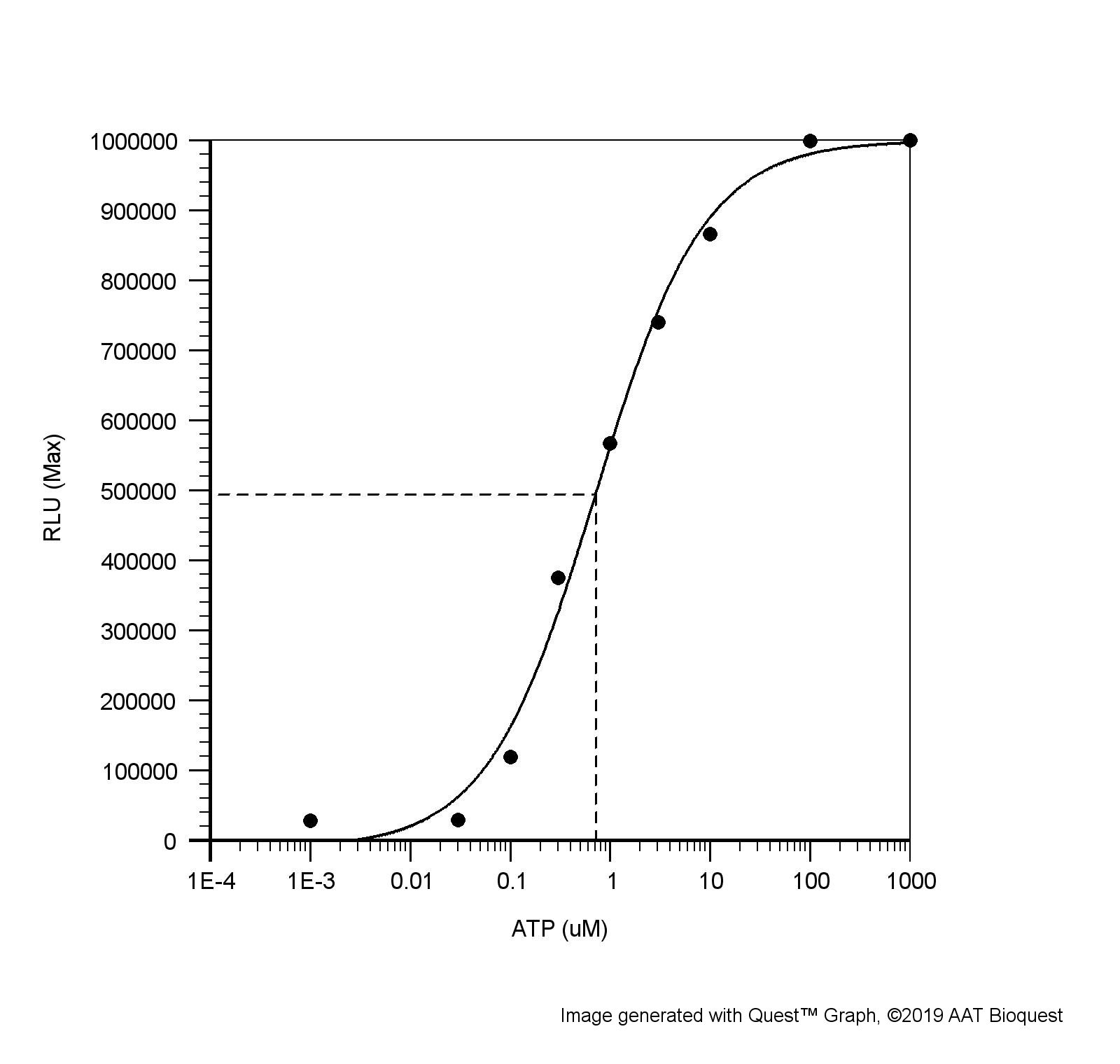
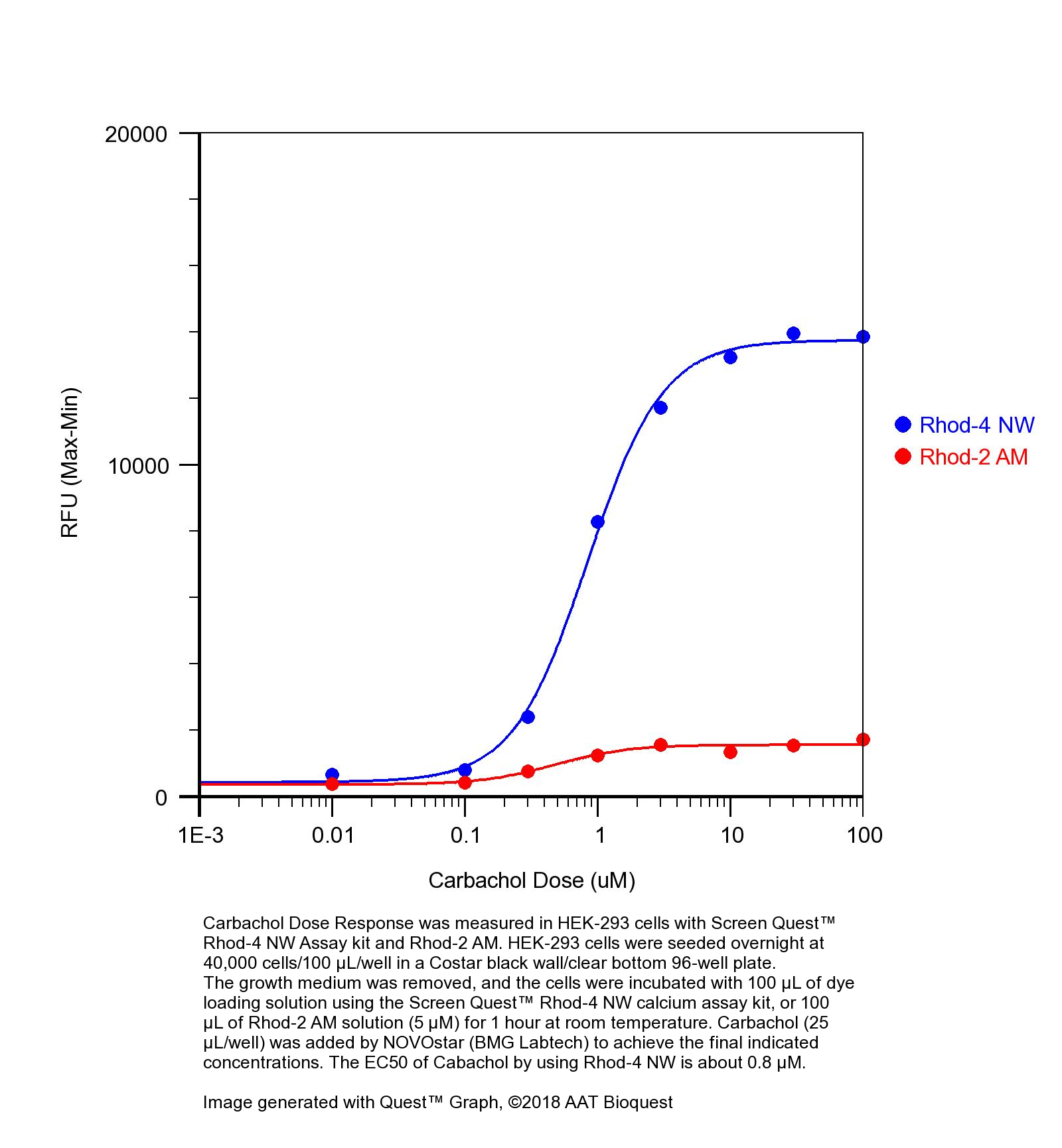
图1.使用Screen Quest Rhod-4 NW分析试剂盒和Rhod-2 AM在HEK-293细胞中测量了卡巴胆碱剂量反应。 将HEK-293细胞以40,000个细胞/ 100 µL /孔在Costar黑色96孔板中过夜接种。 去除生长培养基,并使用Screen Quest Rhod-4 NW钙分析试剂盒将细胞与100 µL染料上样溶液或室温下100 µL Rhod-2 AM溶液(5 µM)孵育1小时。 NOVOstar(BMG Labtech)添加了卡巴胆碱(25 µL /孔)以达到最终指示的浓度。 使用Rhod-4 NW时卡巴胆的EC50约为0.8 µM。


参考文献

Fluorescence absorbance inner-filter decomposition: the role of emission shape on estimates of free Ca(2+) using Rhod-2
Authors: Territo PR, Heil J, Bose S, Evans FJ, Balaban RS.
Journal: Appl Spectrosc (2007): 138

Kinetic characterization of novel NR2B antagonists using fluorescence detection of calcium flux
Authors: Bednar B, Cunningham ME, Kiss L, Cheng G, McCauley JA, Liverton NJ, Koblan KS.
Journal: J Neurosci Methods (2004): 247

Novel fluo-4 analogs for fluorescent calcium measurements
Authors: Martin VV, Beierlein M, Morgan JL, Rothe A, Gee KR.
Journal: Cell Calcium (2004): 509

Protein kinase C and myocardial calcium handling during ischemia and reperfusion: lessons learned using Rhod-2 spectrofluorometry
Authors: Stamm C, del Nido PJ.
Journal: Thorac Cardiovasc Surg (2004): 127

Cytosolic calcium in the ischemic rabbit heart: assessment by pH- and temperature-adjusted rhod-2 spectrofluorometry
Authors: Stamm C, Friehs I, Choi YH, Zurakowski D, McGowan FX, del Nido PJ.
Journal: Cardiovasc Res (2003): 695

Calcium measurements in perfused mouse heart: quantitating fluorescence and absorbance of Rhod-2 by application of photon migration theory
Authors: Du C, MacGowan GA, Farkas DL, Koretsky AP.
Journal: Biophys J (2001): 549

Calibration of the calcium dissociation constant of Rhod(2)in the perfused mouse heart using manganese quenching
Authors: Du C, MacGowan GA, Farkas DL, Koretsky AP.
Journal: Cell Calcium (2001): 217

Changes in mitochondrial Ca2+ detected with Rhod-2 in single frog and mouse skeletal muscle fibres during and after repeated tetanic contractions
Authors: Lannergren J, Westerblad H, Bruton JD.
Journal: J Muscle Res Cell Motil (2001): 265

Rhod-2 based measurements of intracellular calcium in the perfused mouse heart: cellular and subcellular localization and response to positive inotropy
Authors: MacGowan GA, Du C, Glonty V, Suhan JP, Koretsky AP, Farkas DL.
Journal: J Biomed Opt (2001): 23

Mitochondrial free calcium levels (Rhod-2 fluorescence) and ultrastructural alterations in neuronally differentiated PC12 cells during ceramide-dependent cell death
Authors: Muriel MP, Lambeng N, Darios F, Michel PP, Hirsch EC, Agid Y, Ruberg M.
Journal: J Comp Neurol (2000): 297

说明书
Screen Quest 免洗Rhod-4钙检测试剂盒 去除培养基.pdf

Screen Quest 发光法钙离子检测试剂盒 货号36306-AAT Bioquest荧光染料

上海金畔生物科技有限公司代理AAT Bioquest荧光染料全线产品,欢迎访问AAT Bioquest荧光染料官网了解更多信息。

Screen Quest 发光法钙离子检测试剂盒

Screen Quest 发光法钙离子检测试剂盒

货号 36306 存储条件 在零下15度以下保存, 避免光照
规格 100 Plates 价格 32460
Ex (nm) 437 Em (nm) 466
分子量 溶剂
产品详细介绍

简要概述

钙通量测定法是药物发现中筛选G蛋白偶联受体(GPCR)的首选方法。该试剂盒使用高度钙敏感和膜通透的腔肠素类似物作为用载脂蛋白基因转染细胞的钙指示剂。水母发光蛋白是来自维多利亚水母的钙敏感生物发光蛋白,已广泛用作细胞中的钙指示剂。当与钙离子结合时,水母发光蛋白复合物发出蓝光。发光强度与Ca 2+浓度成正比。我们的腔肠素试剂盒比荧光钙测定试剂盒(如Fluo-4,Fluo-3,Calcium-3和Calcium-4)灵敏得多。该试剂盒提供了一种优化的测定方法,用于检测G蛋白偶联受体(GPCR)和钙通道。该测定可以使用96孔或384孔微孔板进行,并易于适应自动化。金畔生物是AAT Bioquest的中国代理商,为您提供最优质的Screen Quest 发光法钙离子检测试剂盒。

钙离子篇:时间轴式讲解应用于钙离子检测的探针


适用仪器


发光酶标仪  
推荐孔板: 黑色透明
读取模式: 底读模式

产品说明书

样品实验方案

简要概述

  1. 通过去除生长培养基制备细胞
  2. 加入腔肠素装载溶液(对于96孔板为100 µL /孔,对于384孔板为25 µL /孔)
  3. 在室温下孵育3-4小时
  4. 检测水母发光蛋白的发光强度


溶液配制

储备溶液配制

1.腔肠素类似物:将250 µL 100%ETOH(组分B)加到腔肠素类似物(组分A)的小瓶中,并充分混合。 注意:25 µL重组腔肠素类似物对于一块板就足够了。 如果将试管密封,则未使用的腔肠素类似物原液可在<-20℃下保存一个月以上。 避光并避免重复的冻融循环。

2.分析缓冲液(1X):a)对于#36305(10板试剂盒),准备使用1X分析缓冲液(组分C)。b)对于 #36306(100板试剂盒),通过将10 mL的10X分析缓冲液(组分C)稀释到90 mL的HHBS缓冲液(试剂盒中未提供)中,制成1X分析缓冲液,并充分混合。 注意:10 mL 1X分析缓冲液足以用于一块板。 将未使用的1X分析缓冲液存储在4℃下。


工作溶液配制

腔肠素装载溶液:将25 µL ETOH重构的腔肠素类似物(储备溶液制备)添加到10 mL 1X分析缓冲液(储备溶液制备)中,并充分混合。 注意:该工作溶液在室温下至少可稳定2小时,并避免光照。


实验步骤

1.从细胞板中去除生长培养基。 注意:重要的是去除生长培养基,以最大程度地减少化合物对血清或培养基的干扰。 注意:将细胞在含0.5-1%FBS的生长培养基中培养,以避免去除培养基的步骤。 在这种情况下,需要在1X缓冲液中装载2X腔肠素。

2.将100 µL /孔(96孔板)或25 µL /孔(384孔板)的腔肠素加载溶液添加到细胞板中。

3.在避光的条件下,于室温下孵育腔肠素装载板3-4小时。

4.用HHBS或所需的缓冲液准备复合板。

5.通过使用具有光电倍增管的封闭腔室的光子检测系统,检测水母发光蛋白的发光强度。 仪器必须完全排除外部光线。


图示

Screen Quest 发光法钙离子检测试剂盒   货号36306

图1. CHO-aeq细胞上的ATP剂量反应。 用载脂蛋白精稳定转染的CHO细胞以50,000个细胞/ 100 µL /孔接种在Costar白色96孔板中过夜。 除去生长培养基,并使用Screen Quest 腔肠素钙测定试剂盒将细胞与100 µL染料上载溶液一起在室温下孵育3小时,并避光。 由NOVOstar(BMG Labtech)添加ATP(25 µL /孔),以达到最终指示的浓度。 ATP的EC50约为0.8 µM。


相关产品

Bioluminescence Resonance Energy Transfer (BRET) Assay for Determination of Molecular Interactions in Living Cells
Authors: Harikumar, Kaleeckal G and Yan, Yan and Xu, Ting-Hai and Melcher, Karsten and Xu, H Eric and Miller, Laurence J
Journal: (2017)

Intracellular Ca2+ is important for flagellin-triggered defense in Arabidopsis and involves inositol polyphosphate signaling
Authors: Ma, Yi and Zhao, Yichen and Berkowitz, Gerald A
Journal: Journal of Experimental Botany (2017)

Orchestration of salivary secretion mediated by two different dopamine receptors in the blacklegged tick Ixodes scapularis
Authors: Kim, Donghun and Simo, Ladislav and Park, Yoonseong
Journal: Journal of Experimental Biology (2014): 3656–3663

Blue fluorescent protein from the calcium-sensitive photoprotein aequorin: catalytic properties for the oxidation of coelenterazine as an oxygenase
Authors: Inouye S, Sasaki S.
Journal: FEBS Lett (2006): 1977

Crystal structure of obelin after Ca2+-triggered bioluminescence suggests neutral coelenteramide as the primary excited state
Authors: Liu ZJ, Stepanyuk GA, Vysotski ES, Lee J, Markova SV, Malikova NP, Wang BC.
Journal: Proc Natl Acad Sci U S A (2006): 2570

Expression, purification and characterization of calcium-triggered luciferin-binding protein of Renilla reniformis
Authors: Inouye S.
Journal: Protein Expr Purif. (2006)

Extended bioluminescence resonance energy transfer (eBRET) for monitoring prolonged protein-protein interactions in live cells
Authors: Pfleger KD, Dromey JR, Dalrymple MB, Lim EM, Thomas WG, Eidne KA.
Journal: Cell Signal (2006): 1664

In vivo testing of Renilla luciferase substrate analogs in an orthotopic murine model of human glioblastoma
Authors: Otto-Duessel M, Khankaldyyan V, Gonzalez-Gomez I, Jensen MC, Laug WE, Rosol M.
Journal: Mol Imaging (2006): 57

Luminescence of imidazo[1,2-a]pyrazin-3(7H)-one compounds
Authors: Teranishi K.
Journal: Bioorg Chem. (2006)

Mitochondrial adaptations within chronically ischemic swine myocardium
Authors: McFalls EO, Sluiter W, Schoonderwoerd K, Manintveld OC, Lamers JM, Bezstarosti K, van Beusekom HM, Sikora J, Ward HB, Merkus D, Duncker DJ.
Journal: J Mol Cell Cardiol (2006): 980

说明书
Screen Quest 发光法钙离子检测试剂盒.pdf

Screen Quest CHO-Gqz嵌合体细胞系 货号38103-AAT Bioquest荧光染料

上海金畔生物科技有限公司代理AAT Bioquest荧光染料全线产品,欢迎访问AAT Bioquest荧光染料官网了解更多信息。

Screen Quest CHO-Gqz嵌合体细胞系

Screen Quest CHO-Gqz嵌合体细胞系

货号 38103 存储条件 在零下80度以下保存
规格 Each 价格 0
Ex (nm) Em (nm)
分子量 溶剂
产品详细介绍

简要概述

产品基本信息

货号:38103

产品名称:Screen Quest CHO-Gqz嵌合体细胞系

规格:Each


适用仪器


其他仪器
FDSS, FLIPR, ViewLux, NOVOStar, ArrayScan, FlexStation, IN Cell Analyzer


产品介绍

Screen Quest 细胞系是一系列细胞,已成功用于药物发现和筛选环境中,用于研究通常不通过细胞内钙偶联的G蛋白偶联受体(GPCR)。它已与FLIPR,FDSS系统有效结合,与许多受体的非Gq偶联成员结合使用,例如趋化因子,5-羟色胺,谷氨酸,多巴胺,阿片样物质,血管加压素以及α和β-肾上腺素能受体家族。超过60%的已知G蛋白偶联受体通过Gq以外的途径发出信号,从而导致细胞内钙增加,并且随着基因组学揭示出更多的GPCR靶标,这种趋势持续增加。 Screen Quest 细胞系用于研究通常不通过细胞内钙偶联的GPCR。 Screen Quest 细胞系基于一系列G蛋白嵌合体,包括混杂的G蛋白Gα16。嵌合体由Gq蛋白复合物的α亚基组成,该复合物的5个羧基末端氨基酸已被其他G蛋白之一(Gαs,Gαi,Gαo或Gαz)取代。这些氨基酸负责受体与其G蛋白的偶联。这些嵌合体与通常通过cAMP途径起作用的特定非Gq偶联受体的共表达可能会导致受体刺激后细胞内钙信号的产生。 Screen Quest CHO-Gqz细胞系是被嵌合Gqzα亚基蛋白稳定转染的CHO-K1细胞。当用作转染表达Gi偶联受体的宿主细胞时,细胞中组成型表达的Gqz蛋白可使通常通过cAMP途径起作用的转染受体与Gq信号转导和动员的细胞内钙偶联。可以使用钙敏感性染料(例如Calbryte 520 AM,Cal-520 AM,Fluo-8 AM或Fluo-4 AM)和免洗的钙试剂盒检测特定配体对这些细胞中特定的非Gq偶联受体的激活。金畔生物是AAT Bioquest的中国代理商,为您提供最优质的Screen Quest CHO-Gqz嵌合体细胞系。 


图示

Screen Quest CHO-Gqz嵌合体细胞系   货号38103

图1.用Cal-520®,AM(Cat#21130)在CHO-Ga16-NOP细胞中测量了伤害感受肽刺激的钙反应。 将CHO-Ga16-NOP细胞以60,000个细胞/ 100 µL /孔在Costar黑色96孔板中接种过夜。 在含有20 mM Hepes缓冲液(HHBS)的Hanks中,将等体积(100 µL)的10 µMCal-520®AM和2 mM丙磺舒在37°C下孵育1小时。 用具有1 mM丙磺舒的HHBS代替Cal-520®AM上样溶液。 通过FlexStation(分子设备)添加了Nociceptin,以达到最终指示的浓度。


参考文献

A cardiac mitochondrial cAMP signaling pathway regulates calcium accumulation, permeability transition and cell death
Authors: Wang Z, Liu D, Varin A, Nicolas V, Courilleau D, Mateo P, Caubere C, Rouet P, Gomez AM, V and ecasteele G, Fischmeister R, Brenner C.
Journal: Cell Death Dis (2016): e2198

Activation of P2X7 and P2Y11 purinergic receptors inhibits migration and normalizes tumor-derived endothelial cells via cAMP signaling
Authors: Avanzato, D and Genova, T and Pla, A Fiorio and Bernardini, M and Bianco, S and Bussolati, B and Mancardi, D and Giraudo, E and Maione, F and Cassoni, P and others
Journal: Scientific Reports (2016)

Changes in the Arabidopsis thaliana Proteome Implicate cAMP in Biotic and Abiotic Stress Responses and Changes in Energy Metabolism
Authors: Alqurashi M, Gehring C, Marondedze C.
Journal: Int J Mol Sci (2016): 852

Odor-induced cAMP production in Drosophila melanogaster olfactory sensory neurons
Authors: Miazzi F, Hansson BS, Wicher D.
Journal: J Exp Biol (2016): 1798

Role of the cAMP Pathway in Glucose and Lipid Metabolism
Authors: Ravnskjaer K, Madiraju A, Montminy M.
Journal: Handb Exp Pharmacol (2016): 29

The pleiotropic role of exchange protein directly activated by cAMP 1 (EPAC1) in cancer: implications for therapeutic intervention
Authors: Almahariq M, Mei FC, Cheng X.
Journal: Acta Biochim Biophys Sin (Shanghai) (2016): 75

cAMP-Induced Histones H3 Dephosphorylation Is Independent of PKA and MAP Kinase Activations and Correlates With mTOR Inactivation
Authors: Rodriguez P, Rojas J.
Journal: J Cell Biochem (2016): 741

A cAMP Biosensor-Based High-Throughput Screening Assay for Identification of Gs-Coupled GPCR Ligands and Phosphodiesterase Inhibitors
Authors: Vedel L, Brauner-Osborne H, Mathiesen JM.
Journal: J Biomol Screen (2015): 849

Cardiac Hypertrophy Is Inhibited by a Local Pool of cAMP Regulated by Phosphodiesterase 2
Authors: Zoccarato A, Surdo NC, Aronsen JM, Fields LA, Mancuso L, Dodoni G, Stangherlin A, Livie C, Jiang H, Sin YY, Gesellchen F, Terrin A, Baillie GS, Nicklin SA, Graham D, Szabo-Fresnais N, Krall J, V and eput F, Movsesian M, Furlan L, Corsetti V, Hamilton G, Lefkimmiatis K, Sjaastad I, Zaccolo M.
Journal: Circ Res (2015): 707

Cardiac cAMP: production, hydrolysis, modulation and detection
Authors: Boularan C, Gales C.
Journal: Front Pharmacol (2015): 203

说明书
Screen Quest CHO-Gqz嵌合体细胞系.pdf

Screen Quest 活细胞cAMP检测(含细胞系) 货号36383-AAT Bioquest荧光染料

上海金畔生物科技有限公司代理AAT Bioquest荧光染料全线产品,欢迎访问AAT Bioquest荧光染料官网了解更多信息。

Screen Quest 活细胞cAMP检测(含细胞系)

Screen Quest 活细胞cAMP检测(含细胞系)

Screen Quest 活细胞cAMP检测(含细胞系)    货号36383 货号 36383 存储条件 在零下15度以下保存, 避免光照
规格 200 Tests 价格 23868
Ex (nm) 490 Em (nm) 520
分子量 溶剂
产品详细介绍

简要概述

产品基本信息

货号:36383

产品名称:Screen Quest cAMP活细胞检测(含细胞系)

规格:200 Tests

储存条件:-15℃避光防潮

保质期:24个月


产品物理化学光谱特性

激发波长(nm):490

发射波长(nm):520


产品介绍

钙通量(通过Gqpathway偶联)测定法是药物发现中筛选GPCR靶标的首选方法。但是,超过60%的已知GPCR通过与cAMP偶联的腺苷酸环化酶活性发出信号。大多数现有的cAMP分析不仅需要细胞裂解,而且缺乏时间和空间分辨率。 Screen Quest 活细胞cAMP检测试剂盒以高通量形式对细胞内cAMP的变化进行实时检测,而无需细胞裂解步骤。该检测法通过包含外源性环状核苷酸门控通道(CNGC)或混杂G蛋白Gα16的细胞系进行。该通道被升高的细胞内cAMP水平激活,导致离子通量和细胞膜去极化,可以用荧光钙(例如Calbryte 520 AM,Cal-520 AM,Fluo-8 AM或Fluo-4 AM和相应的钙离子,免洗洗钙试剂盒)或荧光膜电位染料进行钙信号的检测。 Gα16与特定的非Gq偶联受体的共表达将在受体刺激后导致细胞内钙信号的产生。 Screen Quest 活细胞cAMP检测试剂盒提供了细胞系和试剂,可通过FLIPR,FDSS或其他等效的荧光酶标仪来检测细胞内cAMP的变化,它已成功用于检测Gs和Gi偶联的GPCR活性。


图示

Screen Quest 活细胞cAMP检测(含细胞系)    货号36383

图1. Screen Quest 活细胞cAMP检测原理


参考文献

A cardiac mitochondrial cAMP signaling pathway regulates calcium accumulation, permeability transition and cell death
Authors: Wang Z, Liu D, Varin A, Nicolas V, Courilleau D, Mateo P, Caubere C, Rouet P, Gomez AM, V and ecasteele G, Fischmeister R, Brenner C.
Journal: Cell Death Dis (2016): e2198

Activation of P2X7 and P2Y11 purinergic receptors inhibits migration and normalizes tumor-derived endothelial cells via cAMP signaling
Authors: Avanzato, D and Genova, T and Pla, A Fiorio and Bernardini, M and Bianco, S and Bussolati, B and Mancardi, D and Giraudo, E and Maione, F and Cassoni, P and others
Journal: Scientific Reports (2016)

Changes in the Arabidopsis thaliana Proteome Implicate cAMP in Biotic and Abiotic Stress Responses and Changes in Energy Metabolism
Authors: Alqurashi M, Gehring C, Marondedze C.
Journal: Int J Mol Sci (2016): 852

Odor-induced cAMP production in Drosophila melanogaster olfactory sensory neurons
Authors: Miazzi F, Hansson BS, Wicher D.
Journal: J Exp Biol (2016): 1798

Role of the cAMP Pathway in Glucose and Lipid Metabolism
Authors: Ravnskjaer K, Madiraju A, Montminy M.
Journal: Handb Exp Pharmacol (2016): 29

The pleiotropic role of exchange protein directly activated by cAMP 1 (EPAC1) in cancer: implications for therapeutic intervention
Authors: Almahariq M, Mei FC, Cheng X.
Journal: Acta Biochim Biophys Sin (Shanghai) (2016): 75

cAMP-Induced Histones H3 Dephosphorylation Is Independent of PKA and MAP Kinase Activations and Correlates With mTOR Inactivation
Authors: Rodriguez P, Rojas J.
Journal: J Cell Biochem (2016): 741

A cAMP Biosensor-Based High-Throughput Screening Assay for Identification of Gs-Coupled GPCR Ligands and Phosphodiesterase Inhibitors
Authors: Vedel L, Brauner-Osborne H, Mathiesen JM.
Journal: J Biomol Screen (2015): 849

Cardiac Hypertrophy Is Inhibited by a Local Pool of cAMP Regulated by Phosphodiesterase 2
Authors: Zoccarato A, Surdo NC, Aronsen JM, Fields LA, Mancuso L, Dodoni G, Stangherlin A, Livie C, Jiang H, Sin YY, Gesellchen F, Terrin A, Baillie GS, Nicklin SA, Graham D, Szabo-Fresnais N, Krall J, V and eput F, Movsesian M, Furlan L, Corsetti V, Hamilton G, Lefkimmiatis K, Sjaastad I, Zaccolo M.
Journal: Circ Res (2015): 707

Cardiac cAMP: production, hydrolysis, modulation and detection
Authors: Boularan C, Gales C.
Journal: Front Pharmacol (2015): 203

说明书
Screen Quest 活细胞cAMP检测(含细胞系).pdf

Screen Quest 免洗Fura-2钙检测试剂盒 货号36321-AAT Bioquest荧光染料

上海金畔生物科技有限公司代理AAT Bioquest荧光染料全线产品,欢迎访问AAT Bioquest荧光染料官网了解更多信息。

Screen Quest 免洗Fura-2钙检测试剂盒

Screen Quest 免洗Fura-2钙检测试剂盒

货号 36321 存储条件 在零下15度以下保存, 避免光照
规格 100 Plates 价格 39060
Ex (nm) 336 Em (nm) 505
分子量 溶剂
产品详细介绍

简要概述

钙通量分析是筛选G蛋白偶联受体(GPCR)药物发现的首选方法。该比值钙测定试剂盒允许均匀测量由G蛋白偶联受体或钙通道激活引起的细胞内钙变化。表达通过钙传递信号的GPCR的细胞被Fura-2AM预加载,Fura-2AM可以穿过细胞膜。一旦进入细胞内,亲脂封闭基团Fura-2AM被酯酶裂解,导致带负电荷的荧光染料留在细胞内,其荧光波长在与钙结合时蓝移。当细胞受到激动剂刺激时,受体发出细胞内钙离子释放的信号,从而大大提高Fura-2在短波处的荧光强度。Fura-2的比色特性使该试剂盒成为比单波长Fluo-4更精确测定细胞钙浓度的理想工具。通过单次添加,该分析很容易在高透性环境中进行。该方法可用于96孔或384孔微孔板,且易于自动化。金畔生物是AAT Bioquest的中国代理商,为您提供最优质的Screen Quest 免洗Fura-2钙检测试剂盒。


适用仪器


荧光酶标仪  
激发: 340/380nm
发射: 510nm
cutoff: 470nm
推荐孔板: 黑色透明
读取模式: 底读模式
其他可用仪器:
FDSS, FLIPR, FlexStation

产品说明书

样品实验方案

简要概述

  1. 在生长培养基中准备细胞
  2. 添加Fura-2 AM染料加载溶液(对于96孔板为100 µL /孔,对于384孔板为25 µL /孔)
  3. 在室温下孵育1-2小时
  4. 检测Ex / Em = 340/510 nm和380/510 nm的荧光强度


溶液配制

储备溶液配制

1. Fura-2 AM储备溶液:将200 µL DMSO加入小瓶Fura-2 AM(组分A)中,并充分混合。 注意:20 µL Fura-2 AM储备溶液足以装满一块板。 如果试管密封严密,可将未使用的Fura-2 AM储备溶液分装并在<-20℃下保存超过一个月。 避光并避免重复的冻融循环。

2.分析缓冲液(1X):a)对于#36320(10板试剂盒),通过将9mL HHBS(组分C)添加到10XPluronic®F127 Plus(1 mL,组分B)中来制成1X分析缓冲液,并充分混合。
b)对于#36321(100个板试剂盒),通过向10XPluronic®F127 Plus(10 mL,组分B)中加入90 mL HHBS(未包括)制成1X分析缓冲液。 注意:10 mL 1X分析缓冲液足以用于一块板。 分装并在<-20℃下存储未使用的1X分析缓冲液。 避光并避免重复的冻融循环。


工作溶液配制

Fura-2 AM染料加载溶液:将20 µL Fura-2 AM储备溶液添加到10 mL 1X分析缓冲液中,并充分混合。 注意:该工作溶液在室温下至少可稳定2小时。


实验步骤

1.将100 µL /孔(96孔板)或25 µL /孔(384孔板)的Fura-2 AM染料加载溶液添加到细胞板中。 注意:如果您的化合物干扰血清,则需要用HHBS缓冲液代替生长培养基。

2.将染料加载板在细胞培养箱中孵育1小时,然后在室温下再孵育20分钟。 注意:如果测定需要37℃,请立即进行实验,而无需进一步室温孵育。

3.用HHBS或所需的缓冲液准备复合板。

4.通过监测Ex / Em = 340/510 nm和380/510 nm处的荧光增加来进行钙通量测定。 注意:在实验前进行信号测试非常重要。 不同的仪器具有各自的强度范围。 注意:对于在FDSS上进行的测定,请在仪器上对Fura-2钙测定使用标准滤波器。


图示

Screen Quest 免洗Fura-2钙检测试剂盒   货号36321

图1.用Screen Quest Fura-2 免洗钙含量测定试剂盒测量的CHO-K1细胞中的ATP剂量反应。 将CHO-K1细胞以40,000个细胞/ 100 µL /孔在Costar黑色96孔板中过夜接种。 将细胞与100 µL Screen Quest Fura-2 免洗钙含量测定试剂盒在室温下孵育1小时。 通过FlexStation(分子设备)添加ATP(50 µL /孔)以达到最终指示的浓度。


参考文献

Calcium imaging of cortical neurons using Fura-2 AM
Authors: Barreto-Chang OL, Dolmetsch RE.
Journal: J Vis Exp. (2009)

Measurement of [Ca2+]i in whole cell suspensions using fura-2
Authors: Hirst RA, Harrison C, Hirota K, Lambert DG.
Journal: Methods Mol Biol (2006): 37

Load of calcium probe Fura -2/AM in Escherichia coli cells
Authors: Shao M, Wang HM, Liu ZH, Shen P, Cai RX.
Journal: Wei Sheng Wu Xue Bao (2005): 805

Abnormal spectra alteration observed in Triton calibration method for measuring [Ca2+]i with fluorescence indicator, fura-2
Authors: Xu T, Yang W, Huo XL, Song T.
Journal: J Biochem Biophys Methods (2004): 219

Fluorescence measurements of free [Mg2+] by use of mag-fura 2 in Salmonella enterica
Authors: Froschauer EM, Kolisek M, Dieterich F, Schweigel M, Schweyen RJ.
Journal: FEMS Microbiol Lett (2004): 49

Photonic crystal fibre enables short-wavelength two-photon laser scanning fluorescence microscopy with fura-2
Authors: McConnell G, Riis E.
Journal: Phys Med Biol (2004): 4757

Cytoplasmic calcium measurement in rotavirus enterotoxin-enhanced green fluorescent protein (NSP4-EGFP) expressing cells loaded with Fura-2
Authors: Berkova Z, Morris AP, Estes MK.
Journal: Cell Calcium (2003): 55

AMPA-induced Ca(2+) influx in cultured rat cortical nonpyramidal neurones: pharmacological characterization using fura-2 microfluorimetry
Authors: Fischer W, Franke H, Scheibler P, Allgaier C, Illes P.
Journal: Eur J Pharmacol (2002): 53

Purinergic receptors have different effects in rat exocrine pancreas. Calcium signals monitored by fura-2 using confocal microscopy
Authors: Novak I, Nitschke R, Amstrup J.
Journal: Cell Physiol Biochem (2002): 83

Measurement of [Ca2+]i in whole cell suspensions using fura-2
Authors: Hirst RA, Harrison C, Hirota K, Lambert DG.
Journal: Methods Mol Biol (1999): 31

说明书
Screen Quest 免洗Fura-2钙检测试剂盒.pdf