美国PALL 半封闭药品无菌检测系统

产品名称:美国PALL 半封闭药品无菌检测系统

产品型号:

产品报价:16

产品特点:美国PALL 半封闭药品无菌检测系统 颇尔半封闭式产品的优点:h污染的机会大大低于开放式膜过滤器。h检测一个样品所消耗的仅是一张膜片和配套管路,其他部件均可反复灭菌使用,减少了成本和废弃物对环境造成的污染。h对于抗生素类药品的检测,其培养基的接种量(50ml)仅为全封闭过滤装置的一半。h多种组合形式使中小使用单位以较低的一次性投资即可开展微生物薄膜过滤检测。

美国PALL 半封闭药品无菌检测系统的详细资料:

美国Pall 半封闭药品无菌检测系统 颇尔半封闭式产品的优点:h污染的机会大大低于开放式膜过滤器。h检测一个样品所消耗的仅是一张膜片和配套管路,其他部件均可反复灭菌使用,减少了成本和废弃物对环境造成的污染。h对于抗生素类药品的检测,其培养基的接种量(50ml)仅为全封闭过滤装置的一半。h多种组合形式使中小使用单位以较低的一次性投资即可开展微生物薄膜过滤检测。

产品编号 产品说明 说    明 包 装
15402 多通支架 铝质三通 1/pkg
15403 铝质六通 1/pkg
4247 47mm 磁性过滤漏斗 150ml 容量 1/pkg
4242 300ml 容量 1/pkg
4238 500ml 容量 1/pkg
4241 300ml 容量,带盖 1/pkg
4235 配件 不锈钢支撑网 1/pkg
4246 盖( 仅配300ml 漏斗) 1/pkg
4221 47mm 不锈钢过滤漏斗 100ml 1/pkg
4040 全塑抽滤瓶 1000ml 1/pkg
4402 真空泵保护器 0.45 μm,50mm 3/pkg
13158 无油无尘真空正压双向泵   1/pkg
51147 滤膜镊子   1/pkg
66191 GN-6 单独灭菌膜 0.45 μ47mm 2000/pkg
66585 黑膜,单独灭菌 0.45 μ47mm 200/pkg
66025 吸收垫片 47mm ,灭菌 1000/pkg

Cell Meter 通用Caspase活性荧光法检测试剂盒 绿色荧光 适合于流式细胞检测 货号22821-AAT Bioquest荧光染料

上海金畔生物科技有限公司代理AAT Bioquest荧光染料全线产品,欢迎访问AAT Bioquest荧光染料官网了解更多信息。

Cell Meter 通用Caspase活性荧光法检测试剂盒 绿色荧光 适合于流式细胞检测

Cell Meter 通用Caspase活性荧光法检测试剂盒 绿色荧光 适合于流式细胞检测

Cell Meter 通用Caspase活性荧光法检测试剂盒 绿色荧光 适合于流式细胞检测     货号22821 货号 22821 存储条件 在零下15度以下保存, 避免光照
规格 100 Tests 价格 2604
Ex (nm) 503 Em (nm) 525
分子量 溶剂
产品详细介绍

简要概述

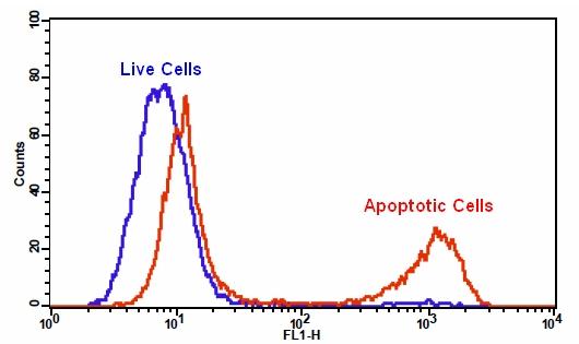
我们的Cell Meter 检测试剂盒是一套用于检测细胞生存力的工具。有多种参数可用于检测细胞活力。该特定试剂盒旨在通过测量活细胞中的通用胱天蛋白酶(胱天蛋白酶-1,-3,-4,-5,-6,-7,-8和-9)激活来检测细胞凋亡。胱天蛋白酶激活被广泛认为是细胞凋亡的可靠指标。大多数胱天蛋白酶具有对肽序列Val-Ala-Asp(VAD)的底物选择性。该试剂盒使用TF2-VAD-FMK作为大多数半胱天冬酶活性的荧光指示剂。TF2-VAD-FMK是细胞渗透性的,无毒的,并且与凋亡细胞中活化的casepase-1,-3,-4,-5,-6,-7,-8和-9不可逆地结合。一旦与胱天蛋白酶结合,绿色荧光试剂将保留在细胞内。结合后阻止了胱天蛋白酶的进一步催化,但不会阻止凋亡的进行。添加到培养基中后的15分钟内,该试剂将开始与活性caspase酶反应。该试剂盒为所有必需成分提供了优化的测定方案。它用于定量凋亡细胞中大多数激活的胱天蛋白酶活性,或筛选胱天蛋白酶抑制剂。绿色荧光标记允许通过流式细胞仪直接检测凋亡细胞中激活的胱天蛋白酶。金畔生物是AAT Bioquest的中国代理商,为您提供最优质的Cell Meter 通用Caspase活性荧光法检测试剂盒。


适用仪器


流式细胞仪  
激发: 488nm激光
发射: 530/30nm滤波片
通道: FITC通道

产品说明书

样品实验方案

简要概述

  1. 用5×10⁵至1×10⁶细胞/ mL的密度制备含测试化合物的细胞
  2. 将0.5 µL细胞溶液中加入1 µL 500X TF2-VAD-FMK
  3. 将细胞在37°C,5%CO2培养箱中孵育1-4小时
  4. 沉淀细胞并将细胞重悬于0.5 mL检测缓冲液或生长培养基中
  5. 使用带有530/30 nm滤光片(FITC通道)的流式细胞仪分析细胞


实验步骤

1.对于每个样品,在0.5 mL温热培养基或自备缓冲液中以5×10⁵至1×10⁶cell/ mL的密度制备细胞。注意:应单独评估每种细胞系,以确定诱导凋亡的最佳细胞密度。

2.用测试化合物处理细胞一段时间,以诱导细胞凋亡,并建立阳性和阴性对照。

3.向处理过的细胞中加入1µL 500X TF2-VAD-FMK(组分A)。

4.将细胞在37°C,5%CO2培养箱中孵育1-4小时。注意:对于贴壁细胞,用0.5 mM EDTA轻轻提起细胞以保持细胞完整,并在与TF2-VAD-FMK孵育之前用含血清的培养基洗涤细胞一次。合适的孵育时间取决于所用的单个细胞类型和细胞浓度。优化每个实验的孵育时间。

5.洗涤并旋转细胞两次。将细胞重悬于0.5 mL分析缓冲液(组分B)或生长培养基中。注意:TF2-VAD-FMK是荧光的;因此,重要的是洗净任何未结合的试剂以除去背景。

6.可选:可以用DNA染色剂标记细胞(例如碘化丙啶或死细胞的7-AAD)。

7.可选:修复细胞。

8.使用带有530/30 nm滤光片的流式细胞仪(FITC通道)检测荧光强度。


相关产品

Angiopoietins Modulate Survival, Migration, and the Components of the Ang-Tie2 Pathway of Chronic Lymphocytic Leukaemia (CLL) Cells In Vitro
Authors: Palma, Luis Mario Aguirre and Flamme, Hanna and Gerke, Iris and Kreuzer, Karl-Anton
Journal: Cancer Microenvironment (2016): 13–26

Hypoxia regulates TRAIL sensitivity of colorectal cancer cells through mitochondrial autophagy.
Authors: Knoll, Gertrud and Bittner, Sebastian and Kurz, Maria and Jantsch, Jonathan and Ehrenschwender, Martin
Journal: Oncotarget (2016)

Promotion of osteointegration under diabetic conditions by tantalum coating-based surface modification on 3-dimensional printed porous titanium implants
Authors: Wang, Lin and Hu, Xiaofan and Ma, Xiangyu and Ma, Zhensheng and Zhang, Yang and Lu, Yizhao and Li, Xiang and Lei, Wei and Feng, Yafei
Journal: Colloids and Surfaces B: Biointerfaces (2016): 440–452

Role of delta-like ligand-4 in chemoresistance against docetaxel in MCF-7 cells
Authors: Wang, Q and Shi, Y and Butler, HJ and Xue, J and Wang, G and Duan, P and Zheng, H
Journal: Human & Experimental Toxicology (2016): 0960327116650006

microRNA-186 inhibits cell proliferation and induces apoptosis in human esophageal squamous cell carcinoma by targeting SKP2
Authors: He, Wei and Feng, Jianfang and Zhang, Yan and Wang, Yuanyuan and Zang, Wenqiao and Zhao, Guoqiang
Journal: Laboratory Investigation (2016): 317–324

Glucose promotes cell proliferation, glucose uptake and invasion in endometrial cancer cells via AMPK/mTOR/S6 and MAPK signaling
Authors: Han, Jianjun and Zhang, Lu and Guo, Hui and Wysham, Weiya Z and Roque, Dario R and Willson, Adam K and Sheng, Xiugui and Zhou, Chunxiao and Bae-Jump, Victoria L
Journal: Gynecologic oncology (2015): 668–675

IL-7 abrogates the immunosuppressive function of human double-negative T cells by activating Akt/mTOR signaling
Authors: Allgäuer, Andrea and Schreiner, Elisabeth and Ferrazzi, Fulvia and Ekici, Arif B and Gerbitz, Armin and Mackensen, Andreas and Völkl, Simon
Journal: The Journal of Immunology (2015): 3139–3148

Insulin improves osteogenesis of titanium implants under diabetic conditions by inhibiting reactive oxygen species overproduction via the PI3K-Akt pathway
Authors: Wang, Lin and Zhao, Xiong and Wei, Bo-yuan and Liu, Yi and Ma, Xiang-yu and Wang, Jian and Cao, Peng-chong and Zhang, Yang and Yan, Ya-bo and Lei, Wei and others
Journal: Biochimie (2015): 85–93

LGL1 modulates proliferation, apoptosis, and migration of human fetal lung fibroblasts
Authors: Zhang, Hui and Sweezey, Neil B and Kaplan, Feige
Journal: American Journal of Physiology-Lung Cellular and Molecular Physiology (2015): L391–L402

t-BHQ Provides Protection against Lead Neurotoxicity via Nrf2/HO-1 Pathway
Authors: Ye, Fang and Li, Xiaoyi and Li, Lili and Yuan, Jing and Chen, Jun
Journal: Oxidative medicine and cellular longevity (2015)

说明书
Cell Meter 通用Caspase活性荧光法检测试剂盒 绿色荧光 适合于流式细胞检测 .pdf

Live或Dead 细胞活性检测试剂盒 绿色/红色双荧光 货号22760-AAT Bioquest荧光染料

上海金畔生物科技有限公司代理AAT Bioquest荧光染料全线产品,欢迎访问AAT Bioquest荧光染料官网了解更多信息。

Live或Dead 细胞活性检测试剂盒 绿色/红色双荧光
Live或Dead 细胞活性检测试剂盒 绿色/红色双荧光

Live或Dead 细胞活性检测试剂盒 绿色/红色双荧光     货号22760 货号 22760 存储条件 在零下15度以下保存, 避免光照
规格 1000 Tests 价格 10548
Ex (nm) 492 Em (nm) 515
分子量 溶剂
产品详细介绍

简要概述

Cell Meter 细胞活性检测试剂盒是美国AAT Bioquest生产的细胞活性检测试剂盒,Cell Meter检测试剂盒是一类用于检测细胞功能的系列工具,包括细胞活性、细胞毒性、细胞凋亡、细胞膜电位以及细胞周期等方面的指标。每种检测方案均能提供不同荧光颜色的检测方案。这些高效的检测方案为从多角度研究细胞功能活动提供了一种十分有效的方法。Cell Meter细胞活力检测试剂盒 *绿色/红色双重荧光* 使用两种非荧光性指示剂:Calcein AM,用于活细胞;一种非细胞渗透性的DNA结合染料,用于细胞膜不完整的死细胞。Calcein AM是一种疏水性复合物,可以轻易地渗透进入完整的活细胞,通过酯酶的水解作用,产生强烈的荧光。通过细胞内酯酶水解非荧光性Calcein AM,形成具有强烈荧光的亲水性Calcein,并且保留在细胞质中。这种酯酶的活性与活细胞数量成比例关系。DNA结合染料极性非常大,不能渗透进入具有完整细胞膜的活细胞,它只有在与死细胞的DNA结合的情况下才发出荧光。生长在培养板中的细胞可以被染料,且在不到两小时内就可以完成定量分析。本检测法比其它活细胞检测法更加强大、稳定。它可以用于许多荧光实验平台的高通量分析中,例如微孔板分析,免疫组化和流式细胞术。本试剂盒提供所有必需组分和最佳检测方案。它适合于增殖型和非增殖型细胞,也可用于悬浮和贴壁细胞。96微孔板中,每孔用试剂100ul,本试剂盒提供的试剂足以进行100次检测;384微孔板中,每孔加试剂25ul,本试剂盒提供的试剂足以进行400次检测。金畔生物是AAT Bioquest的中国代理商,为您提供最优质的Cell Meter 细胞活性检测试剂盒。 


适用仪器


流式细胞仪  
激发: 488nm
发射: 530/30nm, 610/20nm
通道: FITC, PE-Texas Red通道

产品说明书

样品实验方案

简要概述

1.用测试化合物制备细胞
2.加入相同体积的CytoCalcein Green / Propidium Iodide染料加工溶液(100μL/孔/ 96孔板或25μL/孔/ 384孔板)
3.在室温或37°C孵育1小时
4.在强度下监测荧光(底部读取模式)Ex / Em = 490 / 525nm(截止= 515nm)和540 / 620nm(截止= 590nm),带有FITC滤光片(细胞存活)的荧光显微镜和TRITC滤光片 (细胞死亡),或FL1和FL2通道的流式细胞仪。


溶液配制

1.储存溶液配制

        除非另有说明,否则所有未使用的储备溶液应分成一次性等分试样,并在制备后储存在-20°C。 避免反复冻融循环。
CytoCalcein 绿色原液:
将20μLDMSO(组分C)加入到CytoCalcein Green(组分A)的小瓶中并充分混合以制备CytoCalcein Green原液。 避光。 注意:20μL的CytoCalcein Green原液足以用于一个平板。 存放时,密封管。


2.工作溶液配制

将全部内容物(20μL)的CytoCalcein Green储备溶液和20μL碘化丙啶(组分B)加入10mL测定缓冲液(组分C)中并充分混合以制备CytoCalcein Green /碘化丙锭染料 – 工作溶液。 CytoCalcein Green / Propidium Iodide染料加工溶液在室温下稳定至少2小时。 注意:如果CHO细胞等细胞含有导致荧光染料随时间泄漏的有机阴离子转运蛋白,应制备丙磺舒储备溶液,并加入到加样缓冲液中,最终孔内工作浓度范围为1到2.5 mM。 未使用的丙磺舒储备溶液可以在≤-20℃下储存。 由于最佳染色条件可能因细胞类型的不同而异,因此建议单独确定组分A和B的适当浓度。有关细胞样品制备的指南,请点击查看。


操作步骤

1.用酶标仪或荧光显微镜进行细胞活力测定:

1.1根据需要用测试化合物处理细胞。注意:在添加化合物之前不必洗涤细胞。然而,如果测试的化合物对血清敏感,则可在添加化合物之前吸出生长培养基和血清因子。在抽吸后加入100μL/孔/ 96孔板和25μL/孔/ 384孔板的1X Hank盐溶液和20mM Hepes缓冲液(HHBS)或您选择的缓冲液。或者,细胞可以在无血清培养基中生长。

1.2加入100μL/孔(96孔板)或25μL/孔(384孔板)的CytoCalcein TM Green / Propidium Iodide染料加工溶液。

1.3将板在室温或37°C孵育30分钟至1小时,避光。 (孵育时间可以从15分钟到过夜。我们得到了最佳结果,孵育时间少于4小时)。注意:适当的孵育时间取决于所使用的单个细胞类型和细胞浓度。优化每个实验的孵育时间。装载后请勿清洗细胞。对于非粘附细胞,建议在孵育后以800rpm离心细胞板2分钟,然后关闭制动器。

1.4使用荧光酶标仪(底部读取模式)在Ex / Em = 490 / 525nm(截止= 515nm)和Ex / Em = 540 / 620nm(截止= 590nm,细胞死亡)或荧光下监测荧光强度用于活细胞的FITC过滤器或用于死细胞的TRITC过滤器的显微镜。


2.使用流式细胞仪进行细胞活力测定:

2.1用测试化合物处理细胞一段所需的时间。

2.1离心细胞,得到1-5×10⁵个细胞/管。

2.3将细胞重悬于500μL的CytoCalcein™Green / Propidium Iodide染料加工溶液中。

2.4在室温或37°C孵育10至30分钟,避光。

可选:用HHBS或您选择的缓冲液清洗细胞。 将细胞重悬于500μLHHBS中,每管加入1-5×10⁵个细胞。

2.5用流式细胞仪在Ex / Em = 490 / 525nm和Ex / Em = 490 / 620nm(FL1和FL2通道)下监测荧光强度。


数据分析
Live或Dead 细胞活性检测试剂盒 绿色/红色双荧光     货号22760

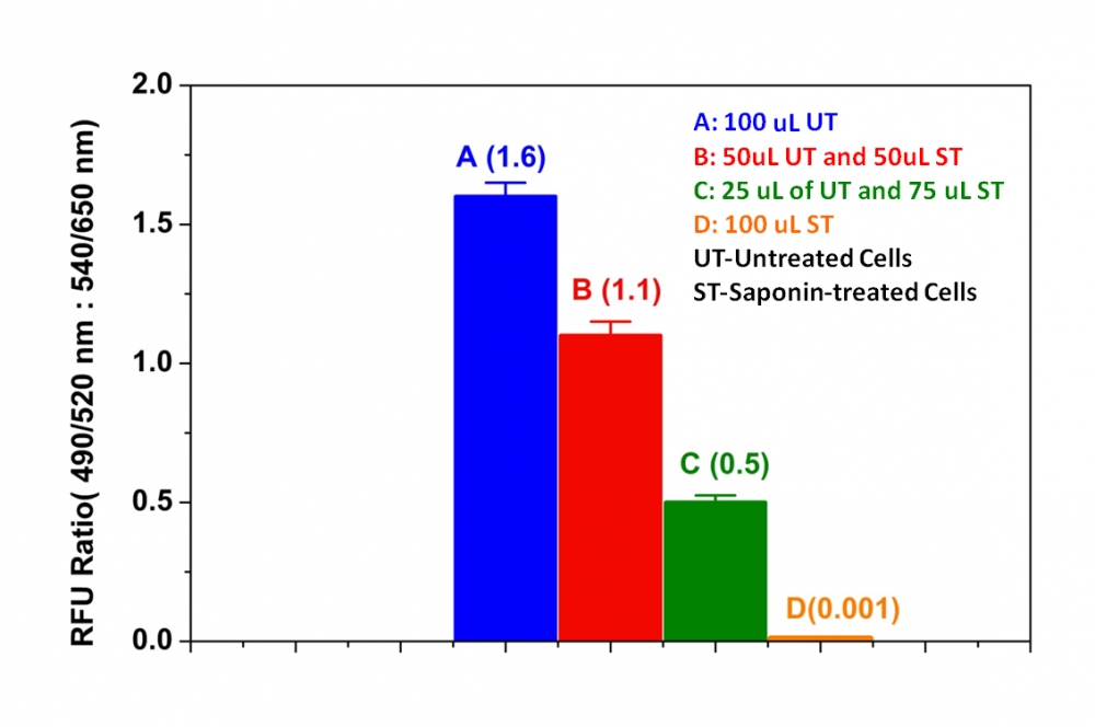
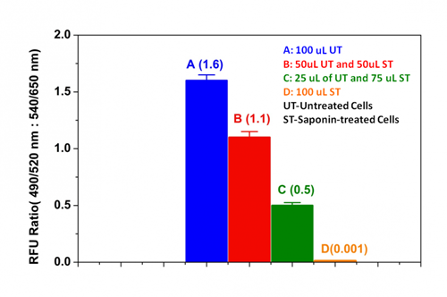
图1. 使用Cell Meter Cell Viability Assay Kit测量Jurkat细胞对皂苷诱导的细胞死亡的影响。 用或不用0.5%皂苷处理2×10 6个细胞/ mL的Jurkat细胞5分钟。 离心细胞,用新鲜培养基替换上清液。 将100uL未处理的细胞(A),各50μL未处理和处理的细胞(B),25uL未处理的和75uL处理的细胞(C)和100uL的0.5%皂苷处理的细胞(D)接种在 96孔黑色墙壁/透明底部聚-D-赖氨酸板。 将细胞与100μL/孔的CytoCalcein Green / Propidium Iodide染料 – 工作溶液在37℃下孵育1小时。 使用NOVOstar仪器(BMG Labtech)在底部读取模式下在Ex / Em = 490 / 525nm和540 / 650nm下测量荧光强度。 如所示(n = 6)显示活细胞和死细胞上490 / 525nm与540 / 650nm荧光强度的比率。


参考文献

Effect of copper nanoparticles on physico-chemical properties of chitosan and gelatin-based scaffold developed for skin tissue engineering application
Authors: Shikha Kumari, Bhisham Narayan Singh, Pradeep Srivastava
Journal: 3 Biotech (2019): 102

Functional imaging of neuronal activity of auditory cortex by using Cal-520 in anesthetized and awake mice
Authors: Jingcheng Li, Jianxiong Zhang, Meng Wang, Junxia Pan, Xiaowei Chen, Xiang Liao
Journal: Biomedical Optics Express (2017): 2599–2610

NINJ2–A novel regulator of endothelial inflammation and activation
Authors: Jingjing Wang, Jingjing Fa, Pengyun Wang, Xinzhen Jia, Huixin Peng, Jing Chen, Yifan Wang, Chenhui Wang, Qiuyun Chen, Xin Tu
Journal: Cellular Signalling (2017)

Erythropoietin Stimulates Endothelial Progenitor Cells to Induce Endothelialization in an Aneurysm Neck After Coil Embolization by Modulating Vascular Endothelial Growth Factor
Authors: Peixi Liu, Yingjie Zhou, Qingzhu An, Yaying Song, Xi Chen, Guo-Yuan Yang, Wei Zhu
Journal: MEDICINE (2016): 1–8

Flexible Endoscopic Spray Application of Respiratory Epithelial Cells as Platform Technology to Apply Cells in Tubular Organs
Authors: Anja Lena Thiebes, Manuel Armin Reddemann, Johannes Palmer, Reinhold Kneer, Stefan Jockenhoevel, Christian Gabriel Cornelissen
Journal: Tissue Engineering Part C: Methods (2016): 322–331

Influence of hypothermia and subsequent rewarming upon leukocyte-endothelial interactions and expression of Junctional-Adhesion-Molecules A and B
Authors: Nicolai V Bogert, Isabella Werner, Angela Kornberger, Patrick Meybohm, Anton Moritz, Till Keller, Ulrich A Stock, Andres Beiras-Fernandez
Journal: Scientific reports (2016)

Inhibition of ABC transport proteins by oil sands process affected water
Authors: Hattan A Alharbi, David MV Saunders, Ahmed Al-Mousa, Jane Alcorn, Alberto S Pereira, Jonathan W Martin, John P Giesy, Steve B Wiseman
Journal: Aquatic Toxicology (2016): 81–88

Rapid generation of collagen-based microtissues to study cell–matrix interactions
Authors: Marie-Elena Brett, Alexandra L Crampton, David K Wood
Journal: Technology (2016): 1–8

Toxicokinetics and toxicodynamics of chlorpyrifos is altered in embryos of Japanese medaka exposed to oil sands process-affected water: evidence for inhibition of P-glycoprotein
Authors: Hattan A Alharbi, Jane Alcorn, Ahmed Al-Mousa, John P Giesy, Steve B Wiseman
Journal: Journal of Applied Toxicology (2016)

Spraying respiratory epithelial cells to coat tissue-engineered constructs
Authors: Anja Lena Thiebes, Stefanie Albers, Christian Klopsch, Stefan Jockenhoevel, Christian G Cornelissen
Journal: BioResearch open access (2015): 278–287


相关产品

产品名称 货号
Live or Dead 细胞活性检测试剂盒 *红/蓝双色荧光* Cat#22788

说明书
Live或Dead 细胞活性检测试剂盒 绿色/红色双荧光 .pdf
Cell Meter 磷脂酰丝氨酸凋亡检测试剂盒 橙色荧光,适合酶标仪检测 货号22794-AAT Bioquest荧光染料

上海金畔生物科技有限公司代理AAT Bioquest荧光染料全线产品,欢迎访问AAT Bioquest荧光染料官网了解更多信息。

Cell Meter 磷脂酰丝氨酸凋亡检测试剂盒 橙色荧光,适合酶标仪检测

Cell Meter 磷脂酰丝氨酸凋亡检测试剂盒 橙色荧光,适合酶标仪检测

Cell Meter 磷脂酰丝氨酸凋亡检测试剂盒 橙色荧光,适合酶标仪检测     货号22794 货号 22794 存储条件 在2-8度冷藏保存, 避免光照
规格 100 Tests 价格 2604
Ex (nm) 554 Em (nm) 578
分子量 溶剂
产品详细介绍

简要概述

Cell Meter检测试剂盒是一类用于检测细胞功能的系列工具,该特定试剂盒旨在通过测量磷脂酰丝氨酸(PS)的转运来检测细胞凋亡。在细胞凋亡中,PS转移到质膜的外部小叶。磷脂酰丝氨酸在细胞表面的出现是细胞凋亡的初始/中间阶段的通用指标,可以在观察形态变化之前进行检测。该试剂盒使用我们专有的橙色荧光Apopxin PS传感器,该传感器特异性结合PS,其亲和力远高于Annexin V(Kd <10 nM)。该试剂盒中使用的PS传感器与膜PS结合后具有橙色荧光。它的光谱性质几乎与Cy3®或AlexaFluor®555的光谱性质相同,使其易于用配备有Cy3®或AlexaFluor®555光源和滤光片的普通荧光仪器(Cy3®或AlexaFluor®555分别是GE Healthcare和Invitrogen的商标)进行实验。由于它对PS的亲和力大大增强,因此该试剂盒比仅与显微镜或流式细胞仪平台一起使用的其他基于膜联蛋白V的商业化凋亡试剂盒更为强大。除显微镜和流式细胞仪平台外,该试剂盒还可与荧光酶标仪一起使用。

点击查看细胞样品配制


适用仪器


荧光酶标仪  
激发: 540nm
发射: 590nm
cutoff: 570nm
推荐孔板: 黑色透明
读取模式: 底读模式

产品说明书

样品实验方案

简要概述

  1. 用测试化合物制备细胞(100 µL /孔/ 96孔板或25 µL /孔/ 384孔板)
  2. 加入等量的Apopxin Orange工作溶液
  3. 在室温下孵育1小时
  4. 在Ex / Em = 540/590 nm(截止= 570 nm)或使用Cy3滤光片的荧光显微镜下检测荧光强度(底部读取模式)


溶液配制

工作溶液配制

将10μL的100X Apopxin 橙色(组分A)添加到1 mL的测定缓冲液(组分B)中,并充分混合以制成Apopxin Orange工作溶液。 注意: 100μLApopxin Orange工作溶液足以用于一个孔。现用现配避免长时间放置。


操作步骤

1.通过向PBS或所需的缓冲液中加入10ul /孔(96孔板)或2.5 µL /孔(384孔板)的10X测试化合物储备溶液来处理细胞。 对于空白孔(不含细胞的培养基),添加相同量的化合物缓冲液。

2.将细胞板在5%CO2、37°C的培养箱中孵育(对于喜树碱处理过的Jurkat细胞,需要4-6小时)以诱导凋亡。

3.在每个孔中添加100 µL /孔(96孔板)或25 µL /孔(384孔板)的Apopxin Orange工作溶液。

4.在避光条件下,于室温下孵育细胞板至少1小时。

5.以800 rpm的速度离心细胞板(特别是非贴壁细胞)2分钟(制动)。

6.使用荧光酶标仪(Ext / Em = 540/590 nm(截止= 570 nm))或使用带有Cy3®滤光片的荧光显微镜对图像进行荧光强度检测。


图示

Cell Meter 磷脂酰丝氨酸凋亡检测试剂盒 橙色荧光,适合酶标仪检测     货号22794

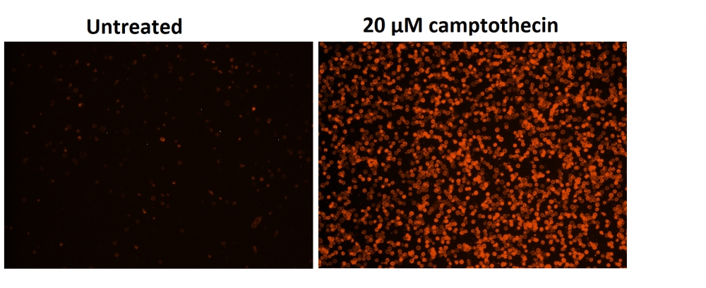
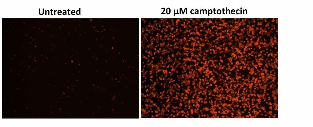
图1.用Cell Meter 磷脂酰丝氨酸细胞凋亡检测试剂盒染色的Costar黑色96孔板中Jurkat细胞的荧光图像。 不使用(左)/使用20 µM喜树碱(右)处理Jurkat细胞5小时。 使用具有通道的荧光显微镜测量荧光强度。


参考文献

Physiological effects of the herbicide glyphosate on the cyanobacterium Microcystis aeruginosa
Authors: Wu, Liang and Qiu, Zhihao and Zhou, Ya and Du, Yuping and Liu, Chaonan and Ye, Jing and Hu, Xiaojun
Journal: Aquatic Toxicology (2016): 72–79

Tumor-selective mitochondrial network collapse induced by atmospheric gas plasma-activated medium.
Authors: Saito, Kosuke and Asai, Tomohiko and Fujiwara, Kyoko and Sahara, Junki and Koguchi, Haruhisa and Fukuda, Noboru and Suzuki-Karasaki, Miki and Soma, Masayoshi and Suzuki-Karasaki, Yoshihiro
Journal: Oncotarget (2016)

Inhibition of malignant phenotypes of human osteosarcoma cells by a gene silencer, a pyrrole–imidazole polyamide, which targets an E-box motif
Authors: Taniguchi, Masashi and Fujiwara, Kyoko and Nakai, Yuji and Ozaki, Toshinori and Koshikawa, Nobuko and Toshio, Kojima and Kataba, Motoaki and Oguni, Asako and Matsuda, Hiroyuki and Yoshida, Yukihiro and others
Journal: FEBS open bio (2014): 328–334

Detection of apoptosis based on the interaction between annexin V and phosphatidylserine
Authors: Liu T, Zhu W, Yang X, Chen L, Yang R, Hua Z, Li G.
Journal: Anal Chem (2009): 2410

Evaluation of cell surface expression of phosphatidylserine in ovarian carcinoma effusions using the annexin-V/7-AAD assay: clinical relevance and comparison with other apoptosis parameters
Authors: Dong HP, Holth A, Kleinberg L, Ruud MG, Elstr and MB, Trope CG, Davidson B, Risberg B.
Journal: Am J Clin Pathol (2009): 756

Mobilization of lysosomal calcium regulates the externalization of phosphatidylserine during apoptosis
Authors: Mirnikjoo B, Balasubramanian K, Schroit AJ.
Journal: J Biol Chem (2009): 6918

Peptidic targeting of phosphatidylserine for the MRI detection of apoptosis in atherosclerotic plaques
Authors: Burtea C, Laurent S, Lancelot E, Ballet S, Murariu O, Rousseaux O, Port M, V and er Elst L, Corot C, Muller RN.
Journal: Mol Pharm (2009): 1903

Suicidal membrane repair regulates phosphatidylserine externalization during apoptosis
Authors: Mirnikjoo B, Balasubramanian K, Schroit AJ.
Journal: J Biol Chem (2009): 22512

Trivalent methylated arsenical-induced phosphatidylserine exposure and apoptosis in platelets may lead to increased thrombus formation
Authors: Bae ON, Lim KM, Noh JY, Chung SM, Kim SH, Chung JH.
Journal: Toxicol Appl Pharmacol (2009): 144

Discovery of a phosphatidylserine-recognizing peptide and its utility in molecular imaging of tumour apoptosis
Authors: Thapa N, Kim S, So IS, Lee BH, Kwon IC, Choi K, Kim IS.
Journal: J Cell Mol Med (2008): 1649

说明书
Cell Meter 磷脂酰丝氨酸凋亡检测试剂盒 橙色荧光,适合酶标仪检测 .pdf

Cell Meter Annexin V凋亡检测试剂盒 橙色荧光 适合流式细胞检测 货号22825-AAT Bioquest荧光染料

上海金畔生物科技有限公司代理AAT Bioquest荧光染料全线产品,欢迎访问AAT Bioquest荧光染料官网了解更多信息。

Cell Meter Annexin V凋亡检测试剂盒 橙色荧光 适合流式细胞检测

Cell Meter Annexin V凋亡检测试剂盒 橙色荧光 适合流式细胞检测

Cell Meter Annexin V凋亡检测试剂盒 橙色荧光 适合流式细胞检测     货号22825 货号 22825 存储条件 在2-8度冷藏保存, 避免光照
规格 100 Tests 价格 2604
Ex (nm) 557 Em (nm) 570
分子量 溶剂
产品详细介绍

简要概述

我们的Cell Meter 检测试剂盒是一套用于检测细胞生存力的工具,有多种参数可用于检测细胞活力。 该特定试剂盒旨在通过测量磷脂酰丝氨酸(PS)的转运来检测细胞凋亡。 在细胞凋亡中,PS转移到质膜的外部小叶。 磷脂酰丝氨酸在细胞表面的出现是细胞凋亡初始/中间阶段的普遍指标,可以在观察形态变化之前进行检测。该试剂盒使用与PS特异性结合的荧光膜联蛋白V。 已经证明膜联蛋白V缀合物选择性结合PS。 使用带有575/26 nm发射滤光片组(PE通道)的流式细胞仪,优化了该特定的测定试剂盒以监测细胞凋亡。金畔生物是AAT Bioquest的中国代理商,为您提供最优质的Cell Meter Annexin V凋亡检测试剂盒。

点击查看光谱


适用仪器


流式细胞仪  
激发: 488或532nm激光
发射: 575/26nm
通道: PE通道

产品说明书

样品实验方案

简要概述

  1. 用测试化合物制备细胞(200 µL /样品)
  2. 添加膜联蛋白V-iFluor 555测定溶液
  3. 在室温下孵育30至60分钟
  4. 使用带有575/26 nm滤光片(PE通道)的流式细胞仪或带有Cy3滤光片组的荧光显微镜分析细胞


实验步骤

1.用膜联蛋白V-iFluor 555准备和孵育细胞:

1.1用测试化合物处理细胞一段时间(对于用星形孢菌素处理过的Jurkat细胞,需要4-6小时)以诱导细胞凋亡。

1.2离心细胞以获得1-5×10⁵细胞/管。

1.3将细胞重悬于200 µL分析缓冲液(组分B)中。

1.4向细胞中加入2 µL Annexin V-iFluor 555(组分A)。

1.5避光保存,于室温下孵育30至60分钟。

1.6在使用流式细胞仪或荧光显微镜分析细胞之前,添加300 µL分析缓冲液(组分B)以增加体积。

1.7使用带有575/26 nm滤光片(PE通道)的流式细胞仪或带有Cy3滤光片组的荧光显微镜检测荧光强度。

2.通过使用流式细胞仪进行分析:

2.1使用带有575/26 nm滤光片的流式细胞仪(PE通道)定量Annexin V- iFluor 555结合物。

3.通过使用荧光显微镜进行分析:

3.1孵育后用移液管吸移细胞悬液,用测定缓冲液冲洗1-2次,然后用测定缓冲液重悬细胞。

3.2将细胞添加到载玻片盖玻片的载玻片上。注意:对于贴壁细胞,建议直接在盖玻片上生长细胞。与Annexin V-iFluor 555孵育后,用测定缓冲液冲洗1-2次,然后将测定缓冲液加到盖玻片上。将玻片上的盖玻片倒置并可视化细胞。与膜联蛋白V-iFluor 555孵育后,还可以将细胞固定在2%甲醛中,并在显微镜下观察。

3.3在荧光显微镜下使用Cy3滤光片组,用Annexin V-iFluor 555分析凋亡细胞。将Nuclear Red DCS1(#17552)添加到细胞后,使用Cy5通道测量细胞活力。质膜上的橙色染色表明膜联蛋白V-iFluor 555与细胞表面的PS结合。


参考文献

In situ polymerizable hydrogel incorporated with specific pathogen-free porcine platelet-rich plasma for the reconstruction of the corneal endothelium
Authors: Lin, Yung-Kai and Sharma, Ruchi and Ma, Hsu and Chen, Wen-Shyan and Yao, Chao-Ling
Journal: Journal of the Taiwan Institute of Chemical Engineers (2017)

Novel regulations of MEF2-A, MEF2-D, and CACNA1S in the functional incompetence of adipose-derived mesenchymal stem cells by induced indoxyl sulfate in chronic kidney disease
Authors: Do, Duyen Thi and Phan, Nam Nhut and Wang, Chih-Yang and Sun, Zhengda and Lin, Yen-Chang
Journal: Cytotechnology (2016): 2589–2604

Shear stress-induced alteration of epithelial organization in human renal tubular cells
Authors: Maggiorani, Damien and Dissard, Romain and Belloy, Marcy and Saulnier-Blache, Jean-Sébastien and Casemayou, Audrey and Ducasse, Laure and Grès, S and ra and Bellière, Julie and Caubet, Cécile and Basc and s, Jean-Loup and others
Journal: PloS one (2015): e0131416

Targeting a G-protein-coupled receptor overexpressed in endocrine tumors by magnetic nanoparticles to induce cell death
Authors: Sanchez, Claire and El Hajj Diab, Darine and Connord, Vincent and Clerc, Pascal and Meunier, Etienne and Pipy, Bernard and Payré, Bruno and Tan, Reasmey P and Gougeon, Michel and Carrey, Julian and others
Journal: Acs Nano (2014): 1350–1363

Glycogen synthase kinase-3 and Omi/HtrA2 induce annexin A2 cleavage followed by cell cycle inhibition and apoptosis
Authors: Wang CY, Lin YS, Su WC, Chen CL, Lin CF.
Journal: Mol Biol Cell (2009): 4153

Gold fluorescent annexin A5 as a novel apoptosis detection tool
Authors: Kurschus FC, Pal PP, Baumler P, Jenne DE, Wiltschi B, Budisa N.
Journal: Cytometry A (2009): 626

Detection of apoptosis induced by new type gosling viral enteritis virus in vitro through fluorescein annexin V-FITC/PI double labeling
Authors: Chen S, Cheng AC, Wang MS, Peng X.
Journal: World J Gastroenterol (2008): 2174

Evaluation of annexin V and Calcein-AM as markers of mononuclear cell apoptosis during human immunodeficiency virus infection
Authors: Palma PF, Baggio GL, Spada C, Silva RD, Ferreira SI, Treitinger A.
Journal: Braz J Infect Dis (2008): 108

Measurement of annexin V uptake and lactadherin labeling for the quantification of apoptosis in adherent Tca8113 and ACC-2 cells
Authors: Hu T, Shi J, Jiao X, Zhou J, Yin X.
Journal: Braz J Med Biol Res (2008): 750

Rhodamine B isothiocyanate doped silica-coated fluorescent nanoparticles (RBITC-DSFNPs)-based bioprobes conjugated to Annexin V for apoptosis detection and imaging
Authors: Shi H, He X, Wang K, Yuan Y, Deng K, Chen J, Tan W.
Journal: Nanomedicine (2007): 266

说明书
Cell Meter Annexin V凋亡检测试剂盒 橙色荧光 适合流式细胞检测 .pdf