Cell Meter 活细胞ATP检测试剂盒 货号23015-AAT Bioquest荧光染料

上海金畔生物科技有限公司代理AAT Bioquest荧光染料全线产品,欢迎访问AAT Bioquest荧光染料官网了解更多信息。

Cell Meter 活细胞ATP检测试剂盒

Cell Meter 活细胞ATP检测试剂盒

Cell Meter 活细胞ATP检测试剂盒    货号23015 货号 23015 存储条件 在零下15度以下保存, 避免光照
规格 100 Tests 价格 4224
Ex (nm) Em (nm)
分子量 溶剂
产品详细介绍

简要概述

三磷酸腺苷(ATP)在细胞能量、代谢调节和细胞信号传导中起着基本作用。它被称为细胞内能量转移的“货币分子单位”,以驱动活细胞中的许多生物过程和化学合成。 ATP也是细胞通讯的信号分子,在DNA和RNA合成中起着重要作用。它位于发生细胞呼吸的线粒体中。 ATP水平可用于测量细胞增殖和细胞周期动态。 Cell Meter 活细胞ATP检测试剂盒使研究人员可以使用检测细胞ATP的渗透型红色荧光成像探针ATP Red 来检测活细胞中的ATP水平。 ATP Red 旨在检测活细胞线粒体中的ATP浓度。该探针与AMP,ADP,CMP,CDP,CTP,UMP,UDP,UTP,GMP,GDP或GTP的交叉反应性最小。


适用仪器


荧光酶标仪  
激发: Cy3/TRITC滤波片组
发射: Cy3/TRITC滤波片组
推荐孔板: 黑色透明孔板

产品说明书

实验方案

概述

在生长培养基中准备细胞
将细胞与ATP Red 工作溶液在37°C下孵育15-30分钟
去掉ATP Red 工作溶液
使用Cy3 / TRITC滤波片组的荧光显微镜进行检测


准备细胞

对于贴壁细胞
在96孔板的生长培养基中以10,000至40,000个细胞/孔/ 90μL平板培养细胞过夜,而对于384孔板则以2,500至10,000个细胞/孔/ 20μL平板培养过夜。

对于悬浮细胞
离心培养液中的细胞,然后将细胞沉淀以50,000-100,000细胞/孔/ 90 µL的浓度悬浮于96孔聚D赖氨酸平板中,或10,000-25,000细胞/孔/ 20 / L以获得384孔中的悬浮液 聚-D赖氨酸板。 实验前,将板以800 rpm的速度离心2分钟。
注意:应该对每个细胞系进行单独评估,以确定最佳细胞密度。


溶液配制

除非另有说明,否则所有未使用的储备溶液应分为一次性使用的等分试样,并在制备后储存在-20°C下。 避免重复冻融循环。

ATP Red 储备溶液配制(500X)
将50 µL DMSO(组分C)添加到小瓶ATP Red (组分A)中,制成500X储备液。
注意:50 µL的500X ATP Red 储备溶液足以容纳一个96孔板。 可以将未使用的ATP Red 储备溶液分装,并在≤-20°C下以较小的等分试样存储。 避光,并避免重复的冻融循环。


ATP Red 工作溶液配制
将5 µL 500X储备溶液(组分A)添加到1 mL细胞培养基中,并充分混合。
注意:ATP Red 探针与我们检测的大多数细胞系的细胞培养基兼容。 可以根据特定的细胞类型修改染色条件。


操作步骤

1.在生长培养基中准备细胞。
2.在细胞板中加入等体积的[100 µL /孔(96孔板)或25 µL /孔(384孔板)]。
注意:ATP Red 的最佳浓度因具体应用而异。
3.在避光的条件下,将细胞在37°C下孵育15-30分钟。
4.从每个孔中除去工作溶液。 用测定缓冲液(组分B)或您选择的缓冲液洗涤细胞两次。
5.使用带有Cy3 / TRITC滤光片组的荧光显微镜观察细胞中的荧光信号。


图示

Cell Meter 活细胞ATP检测试剂盒    货号23015

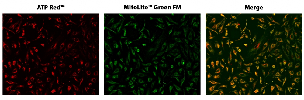
图1. HeLa细胞与ATP Red 和MitoLite Green FM共染色的荧光图像在96孔黑色透明底板。 HeLa细胞用ATP Red 染色15分钟,然后与100 nM MitoLite Green FM(Cat#22695)孵育30分钟。 成像前用测定缓冲液洗涤两次。 ATP Red 和MitoLite ATP Red 信号重叠良好。使用带有Cy3 / TRITC和FITC滤光片的荧光显微镜对细胞成像。


参考文献

Metabolic co-dependence of the oocyte and cumulus cells: essential role in determining oocyte developmental competence.
Authors: Richani, Dulama and Dunning, Kylie R and Thompson, Jeremy G and Gilchrist, Robert B
Journal: Human reproduction update (2021): 27-47

Mitochondrial C3a Receptor Activation in Oxidatively Stressed Epithelial Cells Reduces Mitochondrial Respiration and Metabolism.
Authors: Ishii, Masaaki and Beeson, Gyda and Beeson, Craig and Rohrer, Bärbel
Journal: Frontiers in immunology (2021): 628062

Cell-penetrating and mitochondrion-targeting molecules.
Authors: Appiah Kubi, George and Pei, Dehua
Journal: Methods in enzymology (2020): 311-328

Combinatorial roles of mitochondria and cGMP/PKG pathway in the generation of neuronal free Zn2+ under the presence of nitric oxide.
Authors: Yang, De-Ming and Huang, Chien-Chang and Chang, Yu-Fen
Journal: Journal of the Chinese Medical Association : JCMA (2020): 357-366

Dynamic regulation of subcellular mitochondrial position for localized metabolite levels.
Authors: Alshaabi, Haya and Heininger, Meara and Cunniff, Brian
Journal: Journal of biochemistry (2020): 109-117

Engineering a Reversible Fluorescent Probe for Real-Time Live-Cell Imaging and Quantification of Mitochondrial ATP.
Authors: Ren, Tian-Bing and Wen, Si-Yu and Wang, Lu and Lu, Peng and Xiong, Bin and Yuan, Lin and Zhang, Xiao-Bing
Journal: Analytical chemistry (2020): 4681-4688

Heat stress induces apoptosis through disruption of dynamic mitochondrial networks in dairy cow mammary epithelial cells.
Authors: Chen, Kun-Lin and Wang, Hui-Li and Jiang, Lin-Zheng and Qian, Yong and Yang, Cai-Xia and Chang, Wei-Wei and Zhong, Ji-Feng and Xing, Guang-Dong
Journal: In vitro cellular & developmental biology. Animal (2020): 322-331

Intracellular ATP levels influence cell fates in Dictyostelium discoideum differentiation.
Authors: Hiraoka, Haruka and Nakano, Tadashi and Kuwana, Satoshi and Fukuzawa, Masashi and Hirano, Yasuhiro and Ueda, Masahiro and Haraguchi, Tokuko and Hiraoka, Yasushi
Journal: Genes to cells : devoted to molecular & cellular mechanisms (2020): 312-326

Measurement of ATP concentrations in mitochondria of living cells using luminescence and fluorescence approaches.
Authors: Morciano, Giampaolo and Imamura, Hiromi and Patergnani, Simone and Pedriali, Gaia and Giorgi, Carlotta and Pinton, Paolo
Journal: Methods in cell biology (2020): 199-219

Mitochondria transfer enhances proliferation, migration, and osteogenic differentiation of bone marrow mesenchymal stem cell and promotes bone defect healing.
Authors: Guo, Yusi and Chi, Xiaopei and Wang, Yifan and Heng, Boon Chin and Wei, Yan and Zhang, Xuehui and Zhao, Han and Yin, Ying and Deng, Xuliang
Journal: Stem cell research & therapy (2020): 245

说明书
Cell Meter 活细胞ATP检测试剂盒.pdf

PhosphoWorks 发光测定ATP测定试剂盒*稳定发光* 货号21608-AAT Bioquest荧光染料

上海金畔生物科技有限公司代理AAT Bioquest荧光染料全线产品,欢迎访问AAT Bioquest荧光染料官网了解更多信息。

PhosphoWorks 发光测定ATP测定试剂盒*稳定发光*

PhosphoWorks 发光测定ATP测定试剂盒*稳定发光*

PhosphoWorks 发光测定ATP测定试剂盒*稳定发光*    货号21608 货号 21608 存储条件 在零下15度以下保存, 避免光照
规格 10 Plates 价格 12588
Ex (nm) Em (nm)
分子量 溶剂
产品详细介绍

简要概述

PhosphoWorks 发光测定ATP测定试剂盒*稳定发光*是美国AAT Bioquest生产的用于检测ATP的试剂盒,三磷酸腺苷(ATP)在细胞能量生成,代谢调节和细胞信号传导中起着基本作用。 PhosphoWorks ATP检测试剂盒提供了一种快速,简单且均一的发光检测方法,用于测定哺乳动物细胞中的细胞增殖和细胞毒性。 该测定可以以方便的96孔和384孔微量滴定板形式进行。 此测定法的高灵敏度允许在许多生物系统,环境样品和食品中检测ATP。 该PhosphoWorks ATP检测试剂盒具有长达4小时的稳定发光信号。 它具有稳定的发光,无需混合或分离,并且配方具有最小的动手时间。金畔生物是AAT Bioquest的中国代理商,为您提供最优质的PhosphoWorks 发光测定ATP测定试剂盒*稳定发光*。 


适用仪器


发光酶标仪  
推荐孔板: 白色孔板

产品说明书

样品实验方案

简要概述

1.用测试化合物(100 µL / 96孔板或25 µL / 384孔板)制备细胞(样品)
2.加入等体积的ATP工作溶液(100 µL / 96孔板或25 µL / 384孔板)
3.在室温下孵育10-20分钟
4.检测发光强度


溶液配制

工作溶液配制

1.将整个小瓶10 mL反应缓冲液(组分C)转移到ATP传感器(组分B)中并充分混合。

2.将20 µL ATP监测酶(组分A)添加到组分B + C的瓶子中,并充分混合以制成ATP工作溶液。 注意:避免来自外源性生物来源的潜在ATP污染。

点击查看细胞制备方案


样品操作

1.运行ATP分析:

1.1通过在所需化合物缓冲液中加入10 µL的10X化合物(用于96孔板)或5 µL的5X化合物(用于384孔板)来用测试化合物处理细胞(或样品)。对于空白孔(没有细胞的培养基),添加相应量的化合物缓冲液。

1.2将细胞板在37°C,5%CO2培养箱中孵育所需的时间,例如24、48或96小时。

1.3向每个孔中添加100 µL(96孔板)或25 µL(384孔板)的ATP工作溶液。

1.4在室温下孵育10-20分钟。

1.5使用标准发光计监控发光强度。

2.生成标准ATP校准曲线:

如果需要计算样品中ATP的绝对量,则应与上述测定法一起生成ATP标准曲线。

2.1通过包含不含ATP的样品(作为对照)来测量背景发光,在含0.1%BSA的PBS缓冲液中稀释一系列ATP。注意:通常,ATP浓度从1 nM到10 µM是合适的。

2.2将相同量的稀释的ATP溶液添加到一个空板中(对于96孔板是100 µL,对于384孔板是25 µL)。

2.3加入100 µL /孔(96孔板)或25 µL /孔(384孔板)的ATP工作溶液。

2.4将反应混合物在室温下孵育10至20分钟。

2.5使用标准发光计监控发光强度。

2.6生成ATP标准曲线。


参考文献

High throughput cell-based assay for identification of glycolate oxidase inhibitors as a potential treatment for Primary Hyperoxaluria Type 1
Authors: Mengqiao Wang, Miao Xu, Yan Long, Sonia Fargue, Noel Southall, Xin Hu, John C McKew, Christopher J Danpure, Wei Zheng
Journal: Scientific Reports (2016)

NT1014, a novel biguanide, inhibits ovarian cancer growth in vitro and in vivo
Authors: Lu Zhang, Jianjun Han, Amanda L Jackson, Leslie N Clark, Joshua Kilgore, Hui Guo, Nick Livingston, Kenneth Batchelor, Yajie Yin, Timothy P Gilliam
Journal: Journal of Hematology & Oncology (2016): 91

The Different Effects of Atorvastatin and Pravastatin on Cell Death and PARP Activity in Pancreatic NIT-1 Cells
Authors: Ya-Hui Chen, Yi-Chun Chen, Chin-San Liu, Ming-Chia Hsieh
Journal: Journal of Diabetes Research (2016)

BPA-induced DNA hypermethylation of the master mitochondrial gene PGC-1α contributes to cardiomyopathy in male rats
Authors: Ying Jiang, Wei Xia, Jie Yang, Yingshuang Zhu, Huailong Chang, Juan Liu, Wenqian Huo, Bing Xu, Xi Chen, Yuanyuan Li
Journal: Toxicology (2015): 21–31

Glutamine promotes ovarian cancer cell proliferation through the mTOR/S6 pathway
Authors: Lingqin Yuan, Xiugui Sheng, Adam K Willson, Dario R Roque, Jessica E Stine, Hui Guo, Hannah M Jones, Chunxiao Zhou, Victoria L Bae-Jump
Journal: Endocrine-related cancer (2015): 577–591

JQ1 suppresses tumor growth through downregulating LDHA in ovarian cancer
Authors: Haifeng Qiu, Amanda L Jackson, Joshua E Kilgore, Yan Zhong, Leo Li-Ying Chan, Paola A Gehrig, Chunxiao Zhou, Victoria L Bae-Jump
Journal: Oncotarget (2015): 6915

Loss of histone deacetylase Hdac1 disrupts metabolic processes in intestinal epithelial cells
Authors: Alexis Gonneaud, Naomie Turgeon, Frančois-Michel Boisvert, Frančois Boudreau, Claude Asselin
Journal: FEBS letters (2015): 2776–2783

A neutrophil intrinsic impairment affecting Rab27a and degranulation in cystic fibrosis is corrected by CFTR potentiator therapy
Authors: Kerstin Pohl, Elaine Hayes, Joanne Keenan, Michael Henry, Paula Meleady, Kevin Molloy, Bakr Jundi, David A Bergin, Cormac McCarthy, Oliver J McElvaney
Journal: Blood (2014): 999–1009

Fluorescence lifetime imaging unravels C. trachomatis metabolism and its crosstalk with the host cell
Authors: Márta Szaszák, Philipp Steven, Kensuke Shima, Regina Orzekowsky-Schroder, Gereon Huttmann, Inke R Konig, Werner Solbach, Jan Rupp
Journal: PLoS pathogens (2011): e1002108

G protein coupled receptor kinase 2 interacting protein 1 (GIT1) is a novel regulator of mitochondrial biogenesis in heart
Authors: Jinjiang Pang, Xiangbin Xu, Michael R Getman, Xi Shi, Stephen L Belmonte, Heidi Michaloski, Amy Mohan, Burns C Blaxall, Bradford C Berk
Journal: Journal of molecular and cellular cardiology (2011): 769–776


相关产品

产品名称 货号
PhosphoWorks 发光法ATP检测试剂盒 *明亮发光* Cat#21610
PhosphoWorks 发光法ATP检测试剂盒* DTT – Free* Cat#21612
PhosphoWorks 比色ATP分析试剂盒 Cat#21617

说明书
PhosphoWorks 发光测定ATP测定试剂盒*稳定发光*.pdf

PhosphoWorks 发光法ATP检测试剂盒* DTT – Free* 货号21613-AAT Bioquest荧光染料

上海金畔生物科技有限公司代理AAT Bioquest荧光染料全线产品,欢迎访问AAT Bioquest荧光染料官网了解更多信息。

PhosphoWorks 发光法ATP检测试剂盒* DTT – Free*

PhosphoWorks 发光法ATP检测试剂盒* DTT – Free*

PhosphoWorks 发光法ATP检测试剂盒* DTT - Free*    货号21613 货号 21613 存储条件 在零下15度以下保存, 避免光照
规格 10 Plates 价格 12588
Ex (nm) Em (nm)
分子量 溶剂
产品详细介绍

简要概述

PhosphoWorks 发光法ATP检测试剂盒* DTT – Free*是美国AAT Bioquest生产的用于检测ATP的试剂盒,三磷酸腺苷(ATP)在细胞能量生成,代谢调节和细胞信号传导中起着基本作用。 PhosphoWorks ATP检测试剂盒提供了一种快速,简单且均一的发光检测方法,用于测定哺乳动物细胞中的细胞增殖和细胞毒性。 该测定可以以方便的96孔和384孔微量滴定板形式进行。 此测定法的高灵敏度允许在许多生物系统,环境样品和食品中检测ATP。 此PhosphoWorks ATP检测试剂盒不使用DTT,并且具有长达4小时的稳定发光信号。 它具有稳定的发光,无需混合或分离,并且配方具有最小的动手时间。金畔生物是AAT Bioquest的中国代理商,为您提供最优质的PhosphoWorks 发光法ATP检测试剂盒* DTT – Free*。 


适用仪器


发光酶标仪  
推荐孔板: 白色孔板

产品说明书

操作步骤

简要概述

1.用测试化合物(100μL/ 96孔板或25μL/ 384孔板)制备细胞(样品)
2.加入等体积的ATP工作溶液(100μL/ 96孔板或25μL/ 384孔板)
3.在室温下孵育10-20分钟
4.监测发光强度


制备工作溶液

1.将10 mL反应缓冲液(组分C)转移到ATP传感器(组分B)中并充分混合。

2.将20μLATP监测酶(组分A)加入到组分B + C的瓶中并充分混合以制备ATP工作溶液。 注意:避免来自外源生物来源的潜在ATP污染。

有关细胞样品制备的指南(点击查看)


样品实验方案

1.运行ATP测定:

1.1用测试化合物处理细胞(或样品),在96孔板中加入10μL10X化合物,或在所需化合物缓冲液中加入5μL5X化合物用于384孔板。对于空白孔(没有细胞的培养基),加入相应量的化合物缓冲液。

1.2将细胞板在37℃,5%CO 2培养箱中孵育所需的一段时间,例如24,48或96小时。

1.3向每个孔中加入100μL(96孔板)或25μL(384孔板)的ATP工作溶液。

1.4在室温下孵育10-20分钟。

1.5用标准发光计监测发光强度。


2.生成标准ATP校准曲线:

        如果需要计算样品中ATP的绝对量,则应与上述分析一起生成ATP标准曲线。

2.1通过包含不含ATP的样品(作为对照)在含有0.1%BSA的PBS缓冲液中制备ATP系列稀释液以测量背景发光。注意:通常ATP浓度范围为0.1 nM至1μM是合适的。

2.2将相同量的稀释的ATP溶液加入空板中(对于96孔板为100μL或对于384孔板为25μL)。

2.3加入100μL/孔(96孔板)或25μL/孔(384孔板)的ATP工作溶液。

2.4将反应混合物在室温下孵育10至20分钟。

2.5用标准发光计记录发光强度。

2.6生成ATP标准曲线。


参考文献

High throughput cell-based assay for identification of glycolate oxidase inhibitors as a potential treatment for Primary Hyperoxaluria Type 1
Authors: Mengqiao Wang, Miao Xu, Yan Long, Sonia Fargue, Noel Southall, Xin Hu, John C McKew, Christopher J Danpure, Wei Zheng
Journal: Scientific Reports (2016)

NT1014, a novel biguanide, inhibits ovarian cancer growth in vitro and in vivo
Authors: Lu Zhang, Jianjun Han, Amanda L Jackson, Leslie N Clark, Joshua Kilgore, Hui Guo, Nick Livingston, Kenneth Batchelor, Yajie Yin, Timothy P Gilliam
Journal: Journal of Hematology & Oncology (2016): 91

The Different Effects of Atorvastatin and Pravastatin on Cell Death and PARP Activity in Pancreatic NIT-1 Cells
Authors: Ya-Hui Chen, Yi-Chun Chen, Chin-San Liu, Ming-Chia Hsieh
Journal: Journal of Diabetes Research (2016)

BPA-induced DNA hypermethylation of the master mitochondrial gene PGC-1α contributes to cardiomyopathy in male rats
Authors: Ying Jiang, Wei Xia, Jie Yang, Yingshuang Zhu, Huailong Chang, Juan Liu, Wenqian Huo, Bing Xu, Xi Chen, Yuanyuan Li
Journal: Toxicology (2015): 21–31

Glutamine promotes ovarian cancer cell proliferation through the mTOR/S6 pathway
Authors: Lingqin Yuan, Xiugui Sheng, Adam K Willson, Dario R Roque, Jessica E Stine, Hui Guo, Hannah M Jones, Chunxiao Zhou, Victoria L Bae-Jump
Journal: Endocrine-related cancer (2015): 577–591

JQ1 suppresses tumor growth through downregulating LDHA in ovarian cancer
Authors: Haifeng Qiu, Amanda L Jackson, Joshua E Kilgore, Yan Zhong, Leo Li-Ying Chan, Paola A Gehrig, Chunxiao Zhou, Victoria L Bae-Jump
Journal: Oncotarget (2015): 6915

Loss of histone deacetylase Hdac1 disrupts metabolic processes in intestinal epithelial cells
Authors: Alexis Gonneaud, Naomie Turgeon, Frančois-Michel Boisvert, Frančois Boudreau, Claude Asselin
Journal: FEBS letters (2015): 2776–2783

A neutrophil intrinsic impairment affecting Rab27a and degranulation in cystic fibrosis is corrected by CFTR potentiator therapy
Authors: Kerstin Pohl, Elaine Hayes, Joanne Keenan, Michael Henry, Paula Meleady, Kevin Molloy, Bakr Jundi, David A Bergin, Cormac McCarthy, Oliver J McElvaney
Journal: Blood (2014): 999–1009

Fluorescence lifetime imaging unravels C. trachomatis metabolism and its crosstalk with the host cell
Authors: Márta Szaszák, Philipp Steven, Kensuke Shima, Regina Orzekowsky-Schroder, Gereon Huttmann, Inke R Konig, Werner Solbach, Jan Rupp
Journal: PLoS pathogens (2011): e1002108

G protein coupled receptor kinase 2 interacting protein 1 (GIT1) is a novel regulator of mitochondrial biogenesis in heart
Authors: Jinjiang Pang, Xiangbin Xu, Michael R Getman, Xi Shi, Stephen L Belmonte, Heidi Michaloski, Amy Mohan, Burns C Blaxall, Bradford C Berk
Journal: Journal of molecular and cellular cardiology (2011): 769–776


相关产品

产品名称 货号
PhosphoWorks 发光法ATP检测试剂盒 *温和发光* Cat#21609
PhosphoWorks 发光法ATP检测试剂盒 *明亮发光* Cat#21610
PhosphoWorks 荧光ATP检测试剂盒 Cat#21620

说明书
PhosphoWorks 发光法ATP检测试剂盒* DTT – Free*.pdf

PhosphoWorks 发光法ATP检测试剂盒 *明亮发光* 货号21610-AAT Bioquest荧光染料

上海金畔生物科技有限公司代理AAT Bioquest荧光染料全线产品,欢迎访问AAT Bioquest荧光染料官网了解更多信息。

PhosphoWorks 发光法ATP检测试剂盒 *明亮发光*

PhosphoWorks 发光法ATP检测试剂盒 *明亮发光*

PhosphoWorks 发光法ATP检测试剂盒 *明亮发光*    货号21610 货号 21610 存储条件 在零下15度以下保存, 避免光照
规格 1 Plate 价格 1944
Ex (nm) Em (nm)
分子量 溶剂
产品详细介绍

简要概述

PhosphoWorks 发光法ATP检测试剂盒 *明亮发光*是美国AAT Bioquest生产的用于检测ATP的试剂盒,三磷酸腺苷(ATP)在细胞能量生成,代谢调节和细胞信号传导中起着基本作用。 PhosphoWorks ATP检测试剂盒提供了一种快速,简单且均一的发光检测方法,用于测定哺乳动物细胞中的细胞增殖和细胞毒性。 该测定可以以方便的96孔和384孔微量滴定板形式进行。 此测定法的高灵敏度允许在许多生物系统,环境样品和食品中检测ATP。 该PhosphoWorks ATP分析试剂盒每孔可检测低至10个细胞。 它具有稳定的发光,无需混合或分离,并且配方具有最小的动手时间。金畔生物是AAT Bioquest的中国代理商,为您提供最优质的PhosphoWorks 发光法ATP检测试剂盒 *明亮发光*。 


适用仪器


发光酶标仪  
推荐孔板: 白色孔板

产品说明书

样品实验方案

简要概述

1.用测试化合物(100 µL / 96孔板或25 µL / 384孔板)制备细胞(样品)
2.加入等体积的ATP工作溶液(100 µL / 96孔板或25 µL / 384孔板)
3.在室温下孵育10-20分钟
4.监控发光强度


溶液配制

工作溶液配制

1.将全部反应缓冲液(组分C,10 mL)转移到ATP传感器(组分B)中,并充分混合。

2.将20 µL ATP监测酶(组分A)添加到组分B + C的瓶子中,并充分混合以制成ATP工作溶液。 注意:避免来自外源性生物来源的潜在ATP污染。

点击查看细胞制备方案


样品操作

1.运行ATP分析:

1.1通过在所需化合物缓冲液中加入10 µL的10X化合物(用于96孔板)或5 µL的5X化合物(用于384孔板)来用测试化合物处理细胞(或样品)。对于空白孔(没有细胞的培养基),添加相应量的化合物缓冲液。

1.2将细胞板在37°C,5%CO2培养箱中孵育所需的时间,例如24、48或96小时。

1.3向每个孔中添加100 µL(96孔板)或25 µL(384孔板)的ATP工作溶液。

1.4在室温下孵育10-20分钟。

1.5使用标准发光计监控发光强度。

2.生成标准ATP校准曲线:

如果需要计算样品中ATP的绝对量,则应与上述测定法一起生成ATP标准曲线。

2.1通过包含不含ATP的样品(作为对照)来测量背景发光,在含0.1%BSA的PBS缓冲液中稀释一系列ATP。注意:通常,ATP浓度从1 nM到10 µM是合适的。

2.2将相同量的稀释的ATP溶液添加到一个空板中(对于96孔板是100 µL,对于384孔板是25 µL)。

2.3加入100 µL /孔(96孔板)或25 µL /孔(384孔板)的ATP工作溶液。

2.4将反应混合物在室温下孵育10至20分钟。

2.5使用标准发光计监控发光强度。

2.6生成ATP标准曲线。


参考文献

Hepatitis B Surface Antigen Activates Unfolded Protein Response in Forming Ground Glass Hepatocytes of Chronic Hepatitis B
Authors: Yao Li, Yuchen Xia, Xiaoming Cheng, David E Kleiner, Stephen M Hewitt, Julia Sproch, Tong Li, Hui Zhuang, T Jake Liang
Journal: Viruses (2019): 386

High throughput cell-based assay for identification of glycolate oxidase inhibitors as a potential treatment for Primary Hyperoxaluria Type 1
Authors: Mengqiao Wang, Miao Xu, Yan Long, Sonia Fargue, Noel Southall, Xin Hu, John C McKew, Christopher J Danpure, Wei Zheng
Journal: Scientific Reports (2016)

NT1014, a novel biguanide, inhibits ovarian cancer growth in vitro and in vivo
Authors: Lu Zhang, Jianjun Han, Amanda L Jackson, Leslie N Clark, Joshua Kilgore, Hui Guo, Nick Livingston, Kenneth Batchelor, Yajie Yin, Timothy P Gilliam
Journal: Journal of Hematology & Oncology (2016): 91

The Different Effects of Atorvastatin and Pravastatin on Cell Death and PARP Activity in Pancreatic NIT-1 Cells
Authors: Ya-Hui Chen, Yi-Chun Chen, Chin-San Liu, Ming-Chia Hsieh
Journal: Journal of Diabetes Research (2016)

BPA-induced DNA hypermethylation of the master mitochondrial gene PGC-1α contributes to cardiomyopathy in male rats
Authors: Ying Jiang, Wei Xia, Jie Yang, Yingshuang Zhu, Huailong Chang, Juan Liu, Wenqian Huo, Bing Xu, Xi Chen, Yuanyuan Li
Journal: Toxicology (2015): 21–31

Glutamine promotes ovarian cancer cell proliferation through the mTOR/S6 pathway
Authors: Lingqin Yuan, Xiugui Sheng, Adam K Willson, Dario R Roque, Jessica E Stine, Hui Guo, Hannah M Jones, Chunxiao Zhou, Victoria L Bae-Jump
Journal: Endocrine-related cancer (2015): 577–591

JQ1 suppresses tumor growth through downregulating LDHA in ovarian cancer
Authors: Haifeng Qiu, Amanda L Jackson, Joshua E Kilgore, Yan Zhong, Leo Li-Ying Chan, Paola A Gehrig, Chunxiao Zhou, Victoria L Bae-Jump
Journal: Oncotarget (2015): 6915

Loss of histone deacetylase Hdac1 disrupts metabolic processes in intestinal epithelial cells
Authors: Alexis Gonneaud, Naomie Turgeon, Frančois-Michel Boisvert, Frančois Boudreau, Claude Asselin
Journal: FEBS letters (2015): 2776–2783

A neutrophil intrinsic impairment affecting Rab27a and degranulation in cystic fibrosis is corrected by CFTR potentiator therapy
Authors: Kerstin Pohl, Elaine Hayes, Joanne Keenan, Michael Henry, Paula Meleady, Kevin Molloy, Bakr Jundi, David A Bergin, Cormac McCarthy, Oliver J McElvaney
Journal: Blood (2014): 999–1009

Fluorescence lifetime imaging unravels C. trachomatis metabolism and its crosstalk with the host cell
Authors: Márta Szaszák, Philipp Steven, Kensuke Shima, Regina Orzekowsky-Schroder, Gereon Huttmann, Inke R Konig, Werner Solbach, Jan Rupp
Journal: PLoS pathogens (2011): e1002108


相关产品

产品名称 货号
PhosphoWorks 发光法ATP检测试剂盒 *温和发光* Cat#21609
PhosphoWorks 发光法ATP检测试剂盒* DTT – Free* Cat#21612
PhosphoWorks 比色ATP分析试剂盒 Cat#21617

说明书
PhosphoWorks 发光法ATP检测试剂盒 *明亮发光*.pdf

PhosphoWorks 荧光ATP检测试剂盒 货号21620-AAT Bioquest荧光染料

上海金畔生物科技有限公司代理AAT Bioquest荧光染料全线产品,欢迎访问AAT Bioquest荧光染料官网了解更多信息。

PhosphoWorks 荧光ATP检测试剂盒

PhosphoWorks 荧光ATP检测试剂盒

货号 21620 存储条件 在零下15度以下保存, 避免光照
规格 100 Tests 价格 5244
Ex (nm) 571 Em (nm) 585
分子量 溶剂
产品详细介绍

简要概述

PhosphoWorks 荧光ATP检测试剂盒是美国AAT Bioquest生产的用于检测ATP的试剂盒,三磷酸腺苷(ATP)在细胞能量,代谢调节和细胞信号传导中起着基本作用。它被称为细胞内能量转移的“货币分子单位”,以驱动活细胞中的许多过程和化学合成。 ATP也是细胞通讯的信号分子,在DNA和RNA合成中起重要作用。 AAT Bioquest提供了多种生物发光测定试剂盒,可通过重组萤火虫荧光素酶(目录号21610和21609)确定ATP的纳摩尔(nM)范围。这些试剂盒需要酶标仪,通常用于细胞活力或细胞毒性测定。 PhosphoWorks 荧光ATP分析试剂盒基于一系列ATP诱导的酶偶联反应,产生过氧化氢,该过氧化氢用我们的Amplite Red底物通过分光光度法定量。该测定法可在100 µL反应体积中检测到约0.4 µM ATP,而不受ADP和AMP的干扰。它为测定生物样品中的ATP水平提供了一种强大,简单且方便的测定方法。 PhosphoWorks 荧光ATP分析是我们基于荧光素酶的ATP分析试剂盒的补充。金畔生物是AAT Bioquest的中国代理商,为您提供最优质的PhosphoWorks 荧光ATP检测试剂盒。


适用仪器


荧光酶标仪  
激发: 540nm
发射: 590nm
cutoff: 570nm
推荐孔板: 黑色透明

产品说明书

样品实验方案

简要概述

1.准备ATP工作溶液(50 µL)
2.添加ATP标准液或测试样品(50 µL)
3.在室温下孵育10-30分钟
4.监测Ex / Em = 540/590 nm(截止= 570 nm)的荧光增加


溶液配制

1.制备储备溶液

所有未使用的储备溶液应分为一次性使用的等分试样,并在制备后储存在-20°C下。 避免重复冻融循环。
1.1Amplite 红色底物储备液(200X):
将30 µL DMSO(组分E)加入小瓶Amplite Red Substrate(组分A)中,制成200X Amplite Red Substrate储备液。

1.2ATP标准溶液(10mM):
将0.5 mL的ddH2O加入到ATP标准溶液(组分D)的小瓶中,制成10 mM ATP标准溶液。


2.制备标准溶液

ATP标准

将10 µL的10 mM ATP标准溶液添加到990 µL 1X PBS缓冲液中,以生成100 µM ATP标准溶液(AS7)。 取100 µM ATP标准溶液(AS7)并进行1:3连续稀释,以使用1X PBS缓冲液获得系列稀释的ATP标准溶液(AS6-AS1)。


3.制备工作溶液

3.1将5毫升测定缓冲液(组分C)添加到酶混合瓶(组分B)中,并充分混合。

3.2在Enzyme Mix瓶中加入25 µL 200X AmpliteTM Red Substrate储备液,充分混合以制成APT工作溶液。


样品示例及操作

表1.黑色96孔微孔板中ATP标准品和测试样品的布局。 AS = ATP标准品(AS1-AS7,0.14至100 µM),BL =空白对照,TS =测试样品。

BL BL TS TS
AS1 AS1
AS2 AS2
AS3 AS3    
AS4 AS4    
AS5 AS5    
AS6 AS6    
AS7 AS7    

表2.每个孔的试剂组成

容积 试剂
AS1-AS7 50ul 连续稀释(0.14至100 µM)
BL 50ul 1 X PBS缓冲液
TS 50ul 测试样品

1.根据表1和2中提供的布局,准备ATP标准品(AS),空白对照(BL)和测试样品(TS)。对于384孔板,每孔使用25 µL试剂,而不是50 µL。

2.将50 µL ATP工作溶液添加到ATP标准液,空白对照和测试样品的每个孔中,以使ATP总测定体积为100 µL /孔。 对于384孔板,将25 µL ATP工作溶液添加到每个孔中,总体积为50 µL /孔。

3.避光保存,室温下孵育反应10-30分钟。

4.使用荧光酶标仪在Ex / Em = 540/590 nm(截止= 570 nm)处监视荧光增加。


参考文献

ATP-based cell viability assay is superior to trypan blue exclusion and XTT assay in measuring cytotoxicity of anticancer drugs Taxol and Imatinib, and proteasome inhibitor MG-132 on human hepatoma cell line HepG2
Authors: E. Nowak
Journal: Clin Hemorheol Microcirc (2018): 327-336

High-content image analysis (HCIA) assay has the highest correlation with direct counting cell suspension compared to the ATP, WST-8 and Alamar blue assays for measurement of cytotoxicity
Authors: H. Tahara
Journal: J Pharmacol Toxicol Methods (2017): 92-99

High throughput cell-based assay for identification of glycolate oxidase inhibitors as a potential treatment for Primary Hyperoxaluria Type 1
Authors: Mengqiao Wang, Miao Xu, Yan Long, Sonia Fargue, Noel Southall, Xin Hu, John C McKew, Christopher J Danpure, Wei Zheng
Journal: Scientific Reports (2016)

NT1014, a novel biguanide, inhibits ovarian cancer growth in vitro and in vivo
Authors: Lu Zhang, Jianjun Han, Amanda L Jackson, Leslie N Clark, Joshua Kilgore, Hui Guo, Nick Livingston, Kenneth Batchelor, Yajie Yin, Timothy P Gilliam
Journal: Journal of Hematology & Oncology (2016): 91

The Different Effects of Atorvastatin and Pravastatin on Cell Death and PARP Activity in Pancreatic NIT-1 Cells
Authors: Ya-Hui Chen, Yi-Chun Chen, Chin-San Liu, Ming-Chia Hsieh
Journal: Journal of Diabetes Research (2016)

BPA-induced DNA hypermethylation of the master mitochondrial gene PGC-1α contributes to cardiomyopathy in male rats
Authors: Ying Jiang, Wei Xia, Jie Yang, Yingshuang Zhu, Huailong Chang, Juan Liu, Wenqian Huo, Bing Xu, Xi Chen, Yuanyuan Li
Journal: Toxicology (2015): 21–31

Combination of immunoprecipitation (IP)-ATP_Glo kinase assay and melanogenesis for the assessment of potent and safe PAK1-blockers in cell culture
Authors: B. C. Nguyen
Journal: Drug Discov Ther (2015): 289-95

Glutamine promotes ovarian cancer cell proliferation through the mTOR/S6 pathway
Authors: Lingqin Yuan, Xiugui Sheng, Adam K Willson, Dario R Roque, Jessica E Stine, Hui Guo, Hannah M Jones, Chunxiao Zhou, Victoria L Bae-Jump
Journal: Endocrine-related cancer (2015): 577–591

JQ1 suppresses tumor growth through downregulating LDHA in ovarian cancer
Authors: Haifeng Qiu, Amanda L Jackson, Joshua E Kilgore, Yan Zhong, Leo Li-Ying Chan, Paola A Gehrig, Chunxiao Zhou, Victoria L Bae-Jump
Journal: Oncotarget (2015): 6915

Loss of histone deacetylase Hdac1 disrupts metabolic processes in intestinal epithelial cells
Authors: Alexis Gonneaud, Naomie Turgeon, Frančois-Michel Boisvert, Frančois Boudreau, Claude Asselin
Journal: FEBS letters (2015): 2776–2783


相关产品

产品名称 货号
PhosphoWorks 比色ATP分析试剂盒 Cat#21617
PhosphoWorks 发光法ATP检测试剂盒 *温和发光* Cat#21609
PhosphoWorks 发光法ATP检测试剂盒 *明亮发光* Cat#21610

说明书
PhosphoWorks 荧光ATP检测试剂盒.pdf